

Morphometric analysis of posterior fossa and foramen magnum
- PMID: 23188974
- PMCID: PMC3505313
- DOI: 10.4103/0976-3147.102602
Morphometric analysis of posterior fossa and foramen magnum
Abstract
Introduction: Configuration and size of the foramen magnum and posterior fossa plays an important role in the pathophysiology of the posterior fossa and craniovertebral junction disorders. This study is aimed to find out various dimensions of the foramen magnum and posterior fossa.
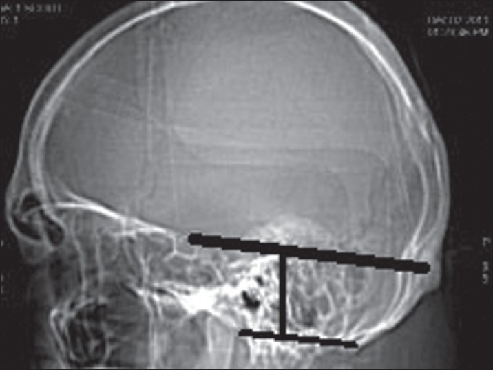
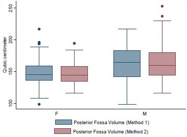
Materials and methods: This is a prospective study of 100 consecutive normal computerized tomography (CT) scans of posterior fossa and 100 dry adult skulls without any bony abnormality. The posterior fossa volume was calculated by abc/2 in method 1 and by advanced work station of CT scan in method 2. Various dimensions of posterior fossa and foramen magnum were also studied.
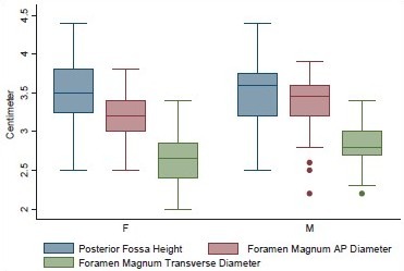
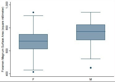
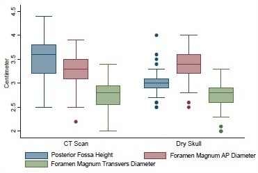
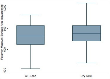
Results: Age ranged from 16 to 89 years with a mean of 51.3 years. Mean height of posterior fossa were 3.01 cm (±0.22) and 3.52 (±0.43) cm in dry skull and CT scan group, respectively (P < 0.0001). Mean volume of posterior fossa were 157.88 (±27.94) cm(3) and 159.58 (±25.73) cm(3) by method 1 and method 2, respectively (P > 0.05). All the dimensions of posterior fossa and foramen magnum were larger in male as compared to female. Mean anteroposterior (AP), transverse diameter and surface area of the foramen magnum were 3.31 (±0.35) cm, 2.76 (±0.31) cm, and 729.15 (±124.87) mm(2), respectively, in CT scan group as compared to 3.41 (±0.29) cm, 2.75 (±0.25) cm, and 747.67 (±108.60) mm(2), respectively, in dry skull group.
Conclusion: Normal values of posterior fossa and foramen magnum could serve as a future reference. Dry skull dimensions could be different from CT scan measurement. More studies are needed as there could be variations in dimensions in different regions in India.
Keywords: Foramen magnum; posterior cranial fossa; skull.
Conflict of interest statement
Figures
Similar articles
-
Morphometric Analysis of Posterior Fossa and Foramen Magnum among Pediatric Age Group 6 to 16 Years.Kathmandu Univ Med J (KUMJ). 2022 Jul-Sep;20(79):342-345. Kathmandu Univ Med J (KUMJ). 2022. PMID: 37042377
-
Computed tomographic study of posterior cranial fossa, foramen magnum, and its surgical implications in Chiari malformations.Asian J Neurosurg. 2017 Jul-Sep;12(3):428-435. doi: 10.4103/1793-5482.175627. Asian J Neurosurg. 2017. PMID: 28761520 Free PMC article.
-
A morphometric analysis of the foramen magnum region as it relates to the transcondylar approach.Acta Neurochir (Wien). 2005 Aug;147(8):889-95. doi: 10.1007/s00701-005-0555-x. Epub 2005 Jun 9. Acta Neurochir (Wien). 2005. PMID: 15924208
-
Chiari 1 deformity in children: etiopathogenesis and radiologic diagnosis.Handb Clin Neurol. 2018;155:25-48. doi: 10.1016/B978-0-444-64189-2.00002-0. Handb Clin Neurol. 2018. PMID: 29891063 Review.
-
[Posterior cranial fossa skull base tumors (clival, foramen magnum, and jugular foramen tumors)].Ryoikibetsu Shokogun Shirizu. 2000;(28 Pt 3):322-7. Ryoikibetsu Shokogun Shirizu. 2000. PMID: 11043259 Review. Japanese. No abstract available.
Cited by
-
The posterior cranial fossa: a comparative MRI-based anatomic study of linear dimensions and volumetry in a homogeneous South Indian population.Surg Radiol Anat. 2015 Oct;37(8):901-12. doi: 10.1007/s00276-015-1434-7. Epub 2015 Jan 28. Surg Radiol Anat. 2015. PMID: 25626883
-
Evaluation of morphological changes in the adult skull with age and sex.J Anat. 2016 Dec;229(6):838-846. doi: 10.1111/joa.12247. Epub 2014 Nov 18. J Anat. 2016. PMID: 25406956 Free PMC article.
-
Binary Logistic Regression Analysis of Foramen Magnum Dimensions for Sex Determination.Anat Res Int. 2015;2015:459428. doi: 10.1155/2015/459428. Epub 2015 Aug 5. Anat Res Int. 2015. PMID: 26346917 Free PMC article.
-
Chiari 1 malformation in patient with Noonan syndrome: A case report and review of literature.Surg Neurol Int. 2025 Apr 11;16:132. doi: 10.25259/SNI_1132_2024. eCollection 2025. Surg Neurol Int. 2025. PMID: 40353171 Free PMC article.
-
Neuraxial dysraphism in EPAS1-associated syndrome due to improper mesenchymal transition.Neurol Genet. 2020 Apr 1;6(3):e414. doi: 10.1212/NXG.0000000000000414. eCollection 2020 Jun. Neurol Genet. 2020. PMID: 32337341 Free PMC article.
References
-
- Bagley CA, Pindrik JA, Bookland MJ, Camara-Quintana JQ, Carson BS. Cervicomedullary decompression for foramen magnum stenosis in achondroplasia. J Neurosurg. 2006;104(3 Suppl):166–72. - PubMed
-
- Dickman C, Spetzler RF, Sonntag VK. Surgery of the craniovertebral junction. 1st ed. New York, NY: Thieme Medical Publishers; 1998.
-
- Wang H, Rosenbaum AE, Reid CS, Zinreich SJ, Pyeritz RE. Pediatric patients with achondroplasia: CT evaluation of the craniocervical junction. Radiology. 1987;164:515–9. - PubMed
-
- Sherekar SK, Yadav YR, Basoor AS, Baghel A, Adam N. Clinical implications of alignment of upper and lower cervical spine. Neurol India. 2006;54:264–7. - PubMed
-
- Kothari RU, Brott T, Broderick JP, Barsan WG, Sauerbeck LR, Zuccarello M, et al. The ABCs of measuring intracerebral hemorrhage volumes. Stroke. 1996;27:1304–5. - PubMed
LinkOut - more resources
Full Text Sources