

Exome analysis identified a novel mutation in the RBP4 gene in a consanguineous pedigree with retinal dystrophy and developmental abnormalities
- PMID: 23189188
- PMCID: PMC3506607
- DOI: 10.1371/journal.pone.0050205
Exome analysis identified a novel mutation in the RBP4 gene in a consanguineous pedigree with retinal dystrophy and developmental abnormalities
Abstract
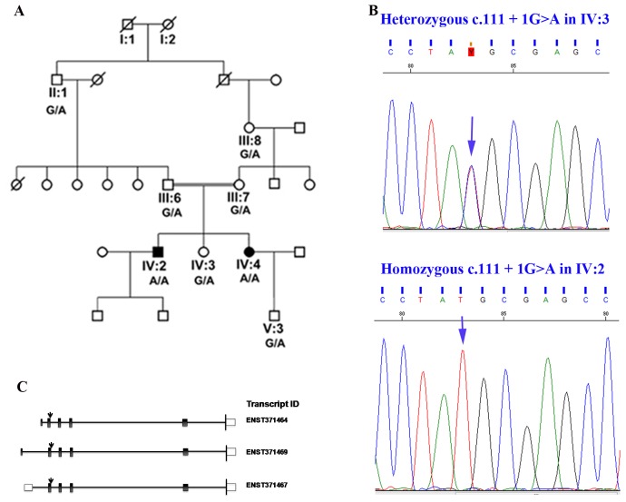
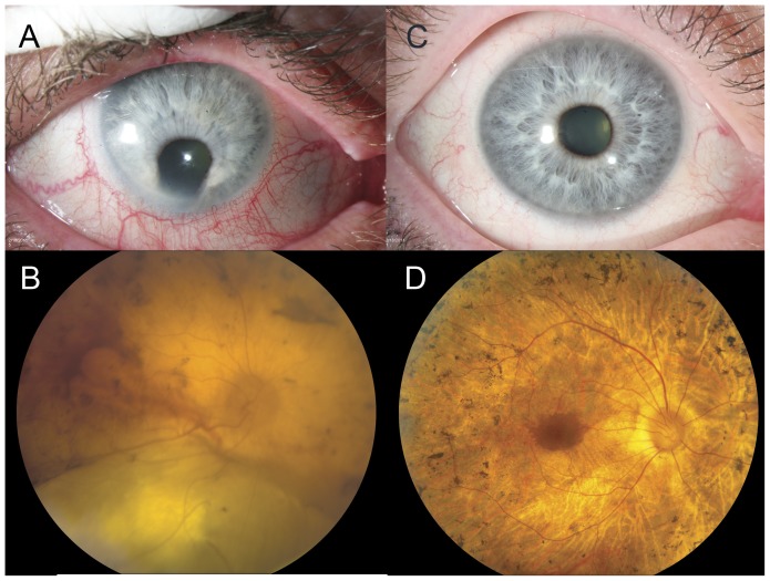
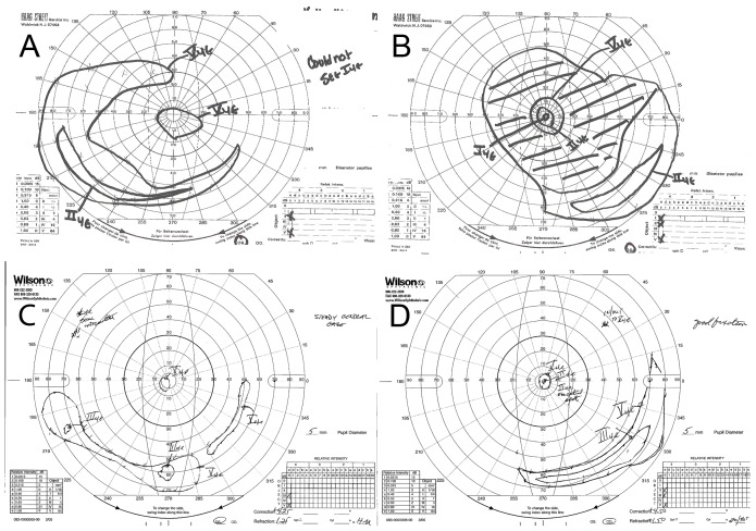
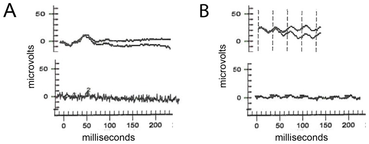
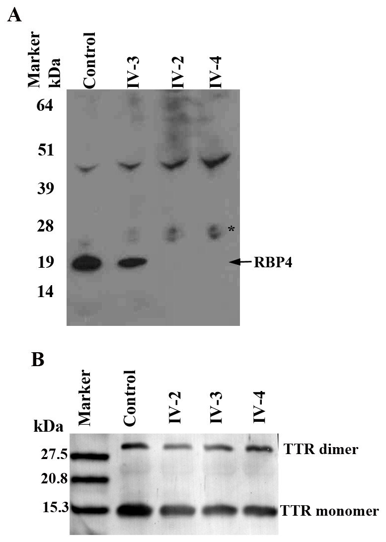
Retinitis Pigmentosa (RP) is a common form of retinal degeneration characterized by photoreceptor degeneration and retinal pigment epithelium (RPE) atrophy causing loss of visual field and acuities. Exome sequencing identified a novel homozygous splice site variant (c.111+1G>A) in the gene encoding retinol binding protein 4 (RBP4). This change segregated with early onset, progressive, and severe autosomal recessive retinitis pigmentosa (arRP) in an eight member consanguineous pedigree of European ancestry. Additionally, one patient exhibited developmental abnormalities including patent ductus arteriosus and chorioretinal and iris colobomas. The second patient developed acne from young age and extending into the 5(th) decade. Both patients had undetectable levels of RBP4 in the serum suggesting that this mutation led to either mRNA or protein instability resulting in a null phenotype. In addition, the patients exhibited severe vitamin A deficiency, and diminished serum retinol levels. Circulating transthyretin levels were normal. This study identifies the RBP4 splice site change as the cause of RP in this pedigree. The presence of developmental abnormalities and severe acne in patients with retinal degeneration may indicate the involvement of genes that regulate vitamin A absorption, transport and metabolism.
Conflict of interest statement
Figures
Similar articles
-
A novel homozygous c.67C>T variant in retinol binding protein 4 (RBP4) associated with retinitis pigmentosa and childhood acne vulgaris.Ophthalmic Genet. 2020 Jun;41(3):288-292. doi: 10.1080/13816810.2020.1755985. Epub 2020 Apr 23. Ophthalmic Genet. 2020. PMID: 32323592
-
Novel C8orf37 Mutations in Patients with Early-onset Retinal Dystrophy, Macular Atrophy, Cataracts, and High Myopia.Ophthalmic Genet. 2016;37(1):68-75. doi: 10.3109/13816810.2014.949380. Epub 2014 Aug 12. Ophthalmic Genet. 2016. PMID: 25113443
-
Vitamin A deficiency due to bi-allelic mutation of RBP4: There's more to it than meets the eye.Ophthalmic Genet. 2017 Sep-Oct;38(5):465-466. doi: 10.1080/13816810.2016.1227453. Epub 2016 Nov 28. Ophthalmic Genet. 2017. PMID: 27892788
-
Identification of a novel mutation in the CDHR1 gene in a family with recessive retinal degeneration.Arch Ophthalmol. 2012 Oct;130(10):1301-8. doi: 10.1001/archophthalmol.2012.1906. Arch Ophthalmol. 2012. PMID: 23044944 Free PMC article.
-
Boucher-Neuhäuser syndrome: cerebellar degeneration, chorioretinal dystrophy and hypogonadotropic hypogonadism: two novel cases and a review of 40 cases from the literature.J Neurol. 2015 Jan;262(1):194-202. doi: 10.1007/s00415-014-7555-9. Epub 2014 Oct 31. J Neurol. 2015. PMID: 25359264 Review.
Cited by
-
Genetic Variation and Mendelian Randomization Approaches.Adv Exp Med Biol. 2022;1390:327-342. doi: 10.1007/978-3-031-11836-4_19. Adv Exp Med Biol. 2022. PMID: 36107328
-
The molecular aspects of absorption and metabolism of carotenoids and retinoids in vertebrates.Biochim Biophys Acta Mol Cell Biol Lipids. 2020 Nov;1865(11):158571. doi: 10.1016/j.bbalip.2019.158571. Epub 2019 Nov 23. Biochim Biophys Acta Mol Cell Biol Lipids. 2020. PMID: 31770587 Free PMC article. Review.
-
An update on the genetics of ocular coloboma.Hum Genet. 2019 Sep;138(8-9):865-880. doi: 10.1007/s00439-019-02019-3. Epub 2019 May 9. Hum Genet. 2019. PMID: 31073883 Review.
-
The Genetics of Acne.Ann Hum Genet. 2025 Sep;89(5):333-341. doi: 10.1111/ahg.70014. Epub 2025 Jul 21. Ann Hum Genet. 2025. PMID: 40689430 Free PMC article. Review.
-
Nutrigenomics: Opportunities & challenges for public health nutrition.Indian J Med Res. 2018 Nov;148(5):632-641. doi: 10.4103/ijmr.IJMR_1738_18. Indian J Med Res. 2018. PMID: 30666988 Free PMC article. Review.
References
-
- Heckenlively JR, Foxman SG, Parelhoff ES (1988) Retinal dystrophy and macular coloboma. Doc Ophthalmol 68: 257–271. - PubMed
-
- Cremers FP, van den Hurk JA, den Hollander AI (2002) Molecular genetics of Leber congenital amaurosis. Hum Mol Genet 11: 1169–1176. - PubMed
-
- Bamshad MJ, Ng SB, Bigham AW, Tabor HK, Emond MJ, et al. (2011) Exome sequencing as a tool for Mendelian disease gene discovery. Nat Rev Genet 12: 745–755. - PubMed
Publication types
MeSH terms
Substances
Grants and funding
LinkOut - more resources
Full Text Sources
Medical
Molecular Biology Databases
Research Materials
Miscellaneous
