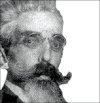
Learning from eponyms: Jose Verocay and Verocay bodies, Antoni A and B areas, Nils Antoni and Schwannomas
- PMID: 23189261
- PMCID: PMC3505436
- DOI: 10.4103/2229-5178.101826
Learning from eponyms: Jose Verocay and Verocay bodies, Antoni A and B areas, Nils Antoni and Schwannomas
Abstract
Schwannomas are benign peripheral nerve sheath neoplasms composed almost entirely of Schwann cells and are diagnosed histopathologically by the presence of singular architectural patterns called Antoni A and Antoni B areas. These were described first in 1920 by the Swedish neurologist Nils Antoni. The Antoni A tissue is highly cellular and made up of palisades of Schwann cell nuclei, a pattern first described in 1910 by the Uruguayan neuro-pathologist Jose Verocay and are known as Verocay bodies. This article describes the structure and appearance of Verocay bodies and Antoni A and B areas with a brief biographical introduction of the men who described these patterns.
Keywords: Antoni A and B areas; Jose Verocay; Nils Antoni; Schwannoma; Verocay bodies.
Conflict of interest statement
Figures


References
-
- Verocay J. [Zur Kennntnis der ‘neurofibrome’] Beitr Pathol Anat. 1910;48:1–69.
-
- Enstable-Puig JF, de Enstable-Puig RF, Haymaker W. [Jose Verocay: Pioneer uruguayan neuropathologist] Arch Fund Roux Ocefa. 1970;4:135–8. - PubMed
-
- Reibel J, Wewer U, Albrechtsen R. The pattern of distribution of laminin in neurogenic tumors, granular cell tumors and nevi of the oral mucosa. Acta Pathol Microbiol Immunol Scand. 1985;93:41–7. - PubMed
-
- Biswas A, Setia N, Bhawan J. Cutaneous neoplasms with prominent Verocay body-like structures: The so called “Rippled Pattern”. Am J Dermatopathol. 2011;33:539–49. quiz 549-50. - PubMed

