. 2012 Mar-Apr;14(2):204-11.
doi: 10.1111/j.1525-142x.2012.00529.x.

Developmental origin of a major difference in sensory patterning between zebrafish and bluefin tuna

Affiliations
Free PMC article

Developmental origin of a major difference in sensory patterning between zebrafish and bluefin tuna

Alain Ghysen et al. Evol Dev. 2012 Mar-Apr.
Free PMC article

Abstract

The posterior lateral line system (PLL) of teleost fish comprises a number of mechanosensory organs arranged in defined patterns on the body surface. Embryonic patterns are largely conserved among teleosts, yet adult patterns are highly diverse. Although changes in pattern modify the perceptual abilities of the system, their developmental origin remains unknown. Here we compare the processes that underlie the formation of the juvenile PLL pattern in Thunnus thynnus, the bluefin tuna, to the processes that were elucidated in Danio rerio, the zebrafish. In both cases, the embryonic PLL comprises five neuromasts regularly spaced along the horizontal myoseptum, but the juvenile PLL comprises four roughly parallel anteroposterior lines in zebrafish, whereas it is a simple dorsally arched line in tuna fish. We examined whether this difference involves evolutionary novelties, and show that the same mechanisms mediate the transition from embryonic to juvenile patterns in both species. We conclude that the marked difference in juveniles depends on a single change (dorsal vs. ventral migration of neuromasts) in the first days of larval life.


Figures

Figure 1
Figure 1
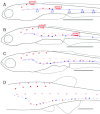
Posterior lateral line (PLL) patterns. Thunnus thynnus PLL at juvenile (A) and embryonic (B) stages. Danio rerio PLL at embryonic (C) and juvenile stages (D). Black dots represent neuromasts, scale bars: 1 mm.
Figure 2
Figure 2
Transition from embryonic to juvenile pattern in zebrafish larvae. (A) The embryonic pattern (blue) is complete at 2 dpf. It comprises five lateral neuromasts (L1–L5) as well as a stripe of interneuromast cells (light blue). (A, B) Migration of post-embryonic primordia primD and prim2, and deposition of their neuromasts (red), extends from 3 to 12 dpf (3–5 mm). In addition to neuromasts, primD and prim2 deposit a discontinuous line of interneuromast cells (pink dashes). (B, C) Ventral migration of neuromasts, and of the stripe of prim1-derived interneuromast cells still attached to neuromasts L1–L5, extends from 4 to 14 dpf (4–6 mm). prim2- and primD-derived interneuromast cells keep their original positions. (C, D) Formation of intercalary neuromasts by local proliferation of prim1-derived interneuromast cells (light blue) extends from 10 to 20 dpf (4.5–7 mm). (D) Formation of intercalary neuromasts by prim2- and primD-derived interneuromast cells (light red) takes place between 15 and 25 dpf (6.5–8 mm). In D, the stripe of prim1-derived interneuromast cells has been omitted for clarity. Scale bars: 1 mm.
Figure 3
Figure 3
Post-embryonic development of Thunnus posterior lateral line (PLL). (A–C) Appearance of a dorsal branch of the PLL nerve (PLLn). (A, B) At 4 days post hatch (corresponding to 6 dpf, 3.6 mm larva) no branch emerges from the PLL; on the next day (4.1 mm), a branch has formed (arrows in C). (D) This branch is lead by a migrating group of mesenchymal cells, prim2 (arrows). (E) The leading axons of the prim2 nerve (prim2n) ramify within prim2. (F) A single confocal frame of the stack used to mount panel E, illustrating the close apposition of peridermal cells (PC), PLL structures, and body muscles (BM). (G) A dorsal branch, primDn, leaves prim2n (arrow in Fig. 2E) and ramifies within a second primordium, primD. (H) prim2n veers away from the horizontal myoseptum (arrows) along a course that corresponds to the stripe of interneuromast cells (dots). The position of prim2 is shown by the arrowhead. (I) At higher magnification, prim2 is seen to migrate just dorsal to the stripe of interneuromast cells (arrowheads). (J) Position of prim2 (arrowhead) at 10dpf (5.2 mm), indicating a rate of migration of about three somites/day. (K) Innervation of prim2-derived proneuromasts by branchlets of prim2n (arrows). (L, M) Proneuromasts assume a rosette-like structure and eventually form pores (arrows in M). (N) prim2-derived neuromasts are (rarely) deposited in close succession, and may even be connected (arrows). prim2-derived neuromasts, and prim1-derived interneuromast cells, keep moving dorsally during larval growth, and progressively separate from prim2n (N). Scale bars: 100 μ.
Figure 4
Figure 4
Post-embryonic hair cell differentiation in Thunnus. (A, B) In a 24 dpf larva (9 mm), phalloidin-labeled apical hair tufts characteristic of differentiated hair cells simultaneously differentiate all along the body, as illustrated here in prim2-derived neuromasts anterior to L1 (A), and between L3 and L4 (B) in the same larva. Most neuromasts comprise two to four hair cells irrespective of their anteroposterior position, and hair cells are invariably polarized along the dorso-ventral axis (insets). Note in A the continuity of embryonic neuromast L1 and interneuromast cells (double arrowhead). Thin arrows in A, C show incipient intercalary neuromasts. (D) Intercalary neuromasts (arrows) are in line with the stripe of interneuromast cells, whereas prim2-derived neuromasts (arrowheads) are dorsal to it. (E) 1.5 cm larva showing the juvenile pattern of a single arched line, dorsal to the horizontal myoseptum (dashed line), and the diminutive dorsal line (arrowheads). (F) Orthogonal polarization of prim2-derived and of intercalary neuromasts. (G, H) prim2-derived neuromasts extend dorsal processes similar to those observed in zebrafish (black and white insets) when neuromasts form stitches. Scale bars: 1 mm in panel E, 100 μ in all other panels, insets in A, B magnified twice.
Figure 5
Figure 5
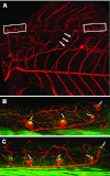
Early differentiation of an ectopic line in a 13 dpf larva. As in normal larvae of this age, primD has formed four to five neuromasts anterior to the dorsal fin (B, boxed in panel A). prim2 has veered dorsally (arrows) instead of following the interneuromast cells past L1, and prim2-derived neuromasts have differentiated precociously (C, boxed in panel A). Arrows point to hair cells kinocilia (red) and stereocilia (green), two clear signs of hair cell differentiation.
Figure 6
Figure 6
Major steps in posterior lateral line larval development in Thunnus. (A) migration paths of prim2 and primD in a 5-mm larva (9 dpf). Both primordia leave small clusters of cells in their wake (red dashes). prim1-derived neuromasts and interneuromasts cells are dark and light blue, respectively. (B) Completion of the prim2 line in a 9-mm larva (20 dpf). (C) Synchronous formation of hair cells in neuromasts of the prim2 line (red dots) in a 10-mm larva (22 dpf). (D) synchronous formation of hair cells in prim1-derived intercalary neuromasts cells (light blue) in a 15-mm larva (25 dpf). Scale bars: 1 mm.

References

    1. Alexandre D, Ghysen A. Somatotopy of the lateral line projection in larval zebrafish. Proc Natl Acad Sci USA. 1999;96:7558–7562. - PMC - PubMed
    1. Aman A, Piotrowski T. Cell migration during morphogenesis. Dev. Biol. 2010;341:20–33. - PubMed
    1. Coombs S, Montgomery JC. The enigmatic lateral line system. In: Fay RR, Popper AN, editors. Comparative Hearing: Fish and Amphibians. New York: Springer Verlag; 1999. pp. 319–362.
    1. Fame RM, Brajon C, Ghysen A. Second-order projection from the posterior lateral line in the early zebrafish brain. Neural Dev. 2006;1:4. - PMC - PubMed
    1. Friedl P, Gilmour D. Collective cell migration in morphogenesis, regeneration and cancer. Nat. Rev. Mol. Cell Biol. 2009;10:445–457. - PubMed

Publication types

MeSH terms

LinkOut - more resources