. 2013 Apr;19(7-8):998-1014.
doi: 10.1089/ten.TEA.2011.0111. Epub 2013 Jan 14.

Three-stage ex vivo expansion of high-ploidy megakaryocytic cells: toward large-scale platelet production

Affiliations

Three-stage ex vivo expansion of high-ploidy megakaryocytic cells: toward large-scale platelet production

Swapna Panuganti et al. Tissue Eng Part A. 2013 Apr.

Abstract

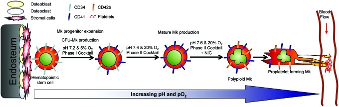
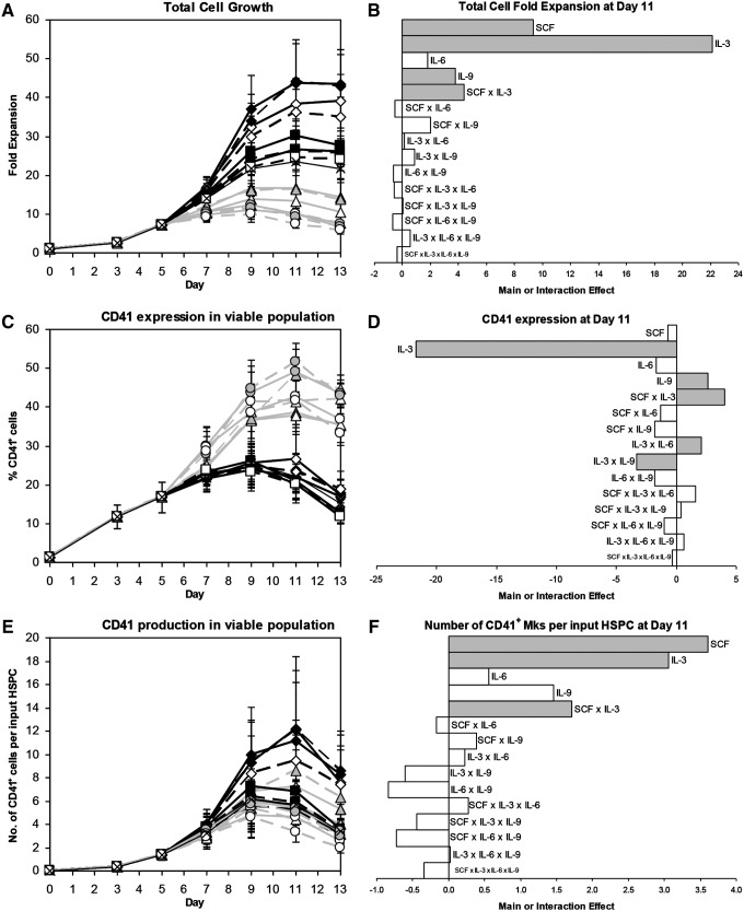
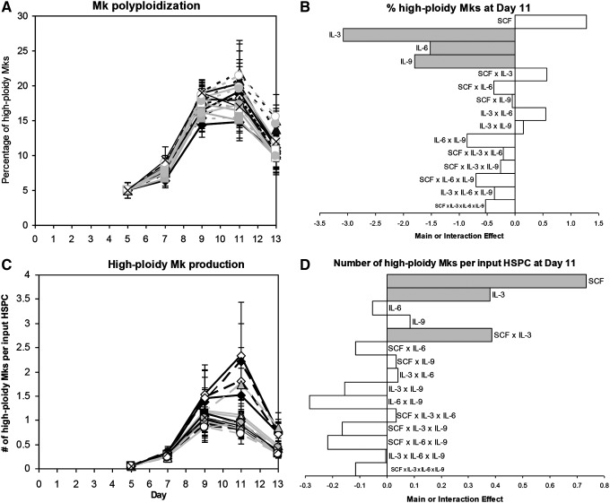
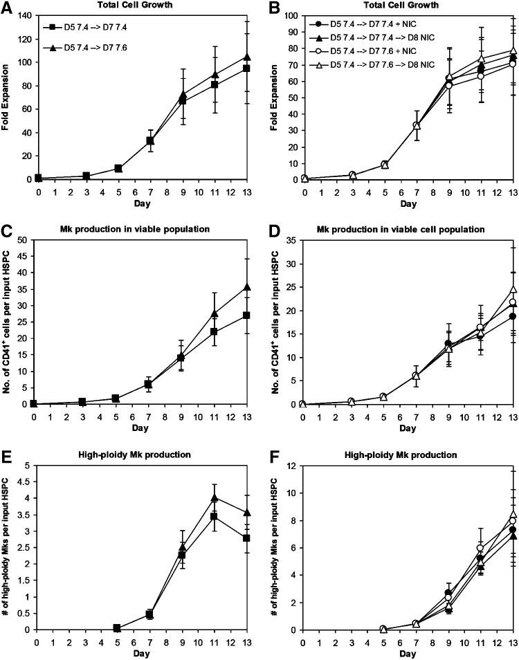
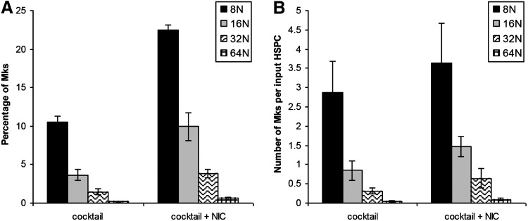
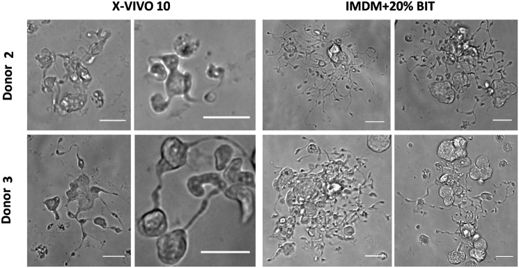
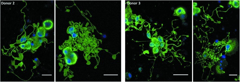
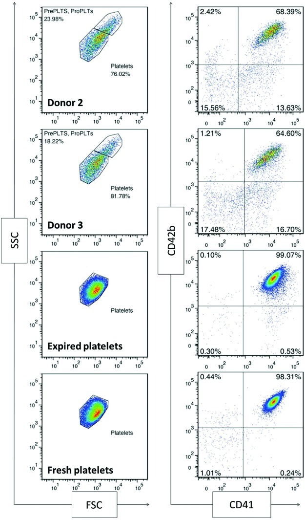
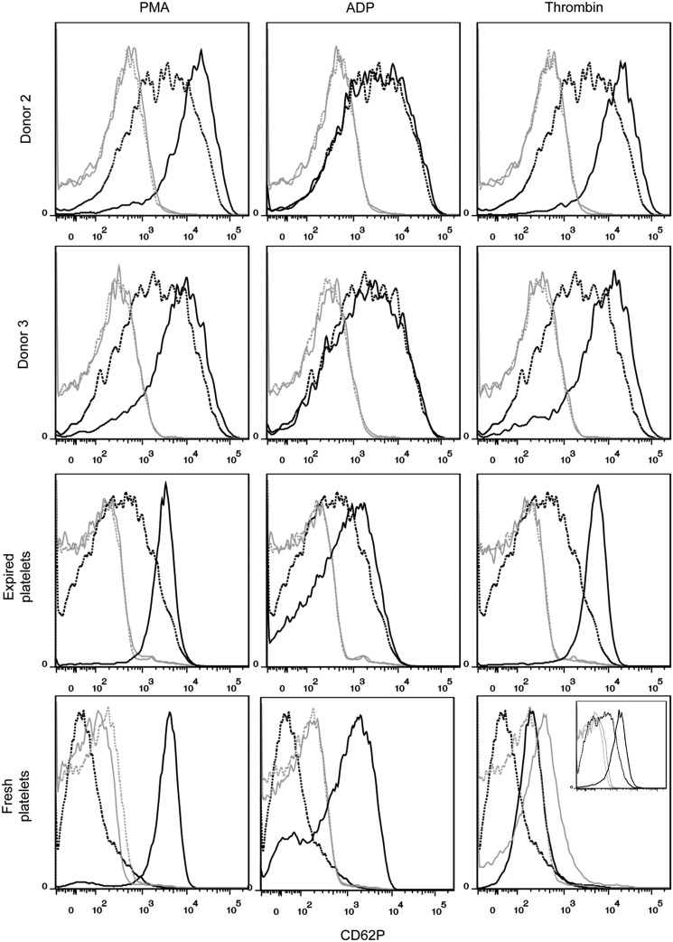
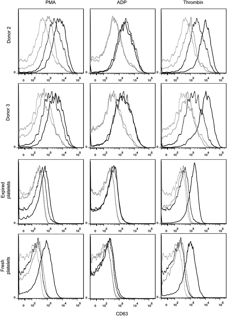
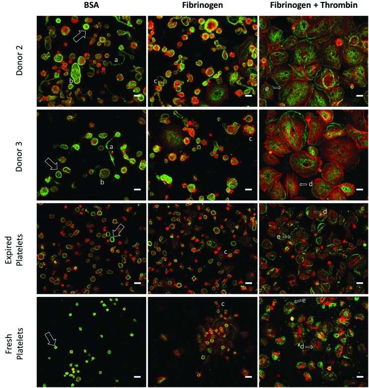
Hematopoietic stem and progenitor cells (HSPCs) have been cultured using a wide variety of cytokines to promote differentiation into megakaryocytic cells (Mks), the precursors to platelets. Greater Mk DNA content, or ploidy, has been correlated with increased platelet release. Gradients of pH, pO2, and signaling factors regulate megakaryopoiesis in the bone marrow niche. In this study, we demonstrate that a 3-phase culture process with increasing pH and pO2 and different cytokine cocktails greatly increases megakaryocyte production. CD34(+) HSPCs were first cultured at 5% O2 and pH 7.2 with a cytokine cocktail previously shown to promote Mk progenitor production. At day 5, cells were shifted to 20% O2 and pH 7.4 and maintained in 1 of 17 cytokine cocktails identified using a 2(4) factorial design of experiments method to evaluate the effects of interleukin (IL)-3, IL-6, IL-9, and high- or low-dose stem cell factor (SCF), in conjunction with thrombopoietin (Tpo) and IL-11, on expansion of mature Mks from progenitors. The combination of Tpo, high-dose SCF, IL-3, IL-9, and IL-11 best promoted Mk expansion. IL-3 greatly increased total cell fold expansion, but this was partially offset by lower Mk purity. IL-9 promoted CD41 and CD42b expression. High-dose (100 ng/mL) SCF increased Mk production and ploidy. Different commercial media and IL-3 sources substantially impacted differentiation, and X-VIVO 10 serum-free media best supported mature Mk expansion. Shifting from pH 7.4 to pH 7.6 at day 7 increased Mk production by 30%. Treatment with nicotinamide at day 7 or day 8 more than doubled the fraction of high-ploidy (>4N) Mks. Ultimately, the 3-phase culture system gave rise to 44.5±8.1 Mks and 8.5±3.1 high-ploidy Mks per input HSPC. Further optimization was required to improve platelet production. Using Iscove's modified Dulbecco's medium (IMDM)+20% BSA, insulin and transferin (BIT) 9500 Serum Substitute greatly improved the frequency and quality of Mk proplatelet extensions without affecting Mk expansion, commitment, or polyploidization in the 3-phase process. Mks cultured in IMDM+20% BIT 9500 gave rise to platelets with functional activity similar to that of fresh platelets from normal donors, as evidenced by basal tubulin distribution and the expression of surface markers and spreading in response to platelet agonists.


Figures

FIG. 1.
FIG. 1.
Schematic of Mk differentiation in vivo and in 3-phase culture. Hematopoietic stem and progenitor cells (HSPCs) reside close to the endosteal bone surface in the niche. During the course of differentiation, megakaryocytic cells (Mks) are exposed to continually increasing pH and pO2 until reaching the sinuses, where platelets are released from proplatelets aided by shear from the blood flow. Expression of CD34 decreases as Mks mature, while expression of CD41 and CD42b increases. Text above the arrows describes the niche-inspired culture system developed to optimize ex vivo Mk production from HSPCs. Color images available online at www.liebertpub.com/tea
FIG. 2.
FIG. 2.
Mk production for 24 factorial design of experiments. Effects of 17 cocktails on (A, B) total cell fold-expansion, (C, D) CD41 expression, and (E, F) Mk yield per HSPC. (A, C, E) Cytokine cocktails are indicated by symbol and line combinations (e.g., open black diamond and solid black line), as shown in Table 1. Data represent the mean±standard error (SEM) for n=5 donors. (B, D, F) Main and interaction cytokine effects at day 11; significant effects (p<0.05) are shaded gray. IL, interleukin; SCF, stem cell factor.
FIG. 3.
FIG. 3.
High-ploidy Mk production for 24 factorial design of experiments. Effects of 17 cocktails on (A, B) the percentage of high-ploidy (>4N) Mks and (C, D) the number of high-ploidy Mks produced per HSPC. (A, C) Cytokine cocktails are indicated by symbol and line combinations (e.g., open black diamond and solid black line), as shown in Table 1. Data represent the mean±SEM for n=5 donors. (B, D) Main and interaction cytokine effects at day 11; significant effects (p<0.05) are shaded gray.
FIG. 4.
FIG. 4.
Increasing pH and adding nicotinamide (NIC) increase high-ploidy Mk production. CD34+ HSPCs were seeded in X-VIVO 10 with cocktail “c” (Table 1) using interleukin (IL)-3 from R&D Systems and maintained at 5% O2 and pH 7.2. On day 5, cells were washed, resuspended at pH 7.4/20% O2, and supplemented with cocktail 3. (A, C, E) On day 7, cells were washed and resuspended in media supplemented with cocktail 3 and adjusted to pH 7.4 (squares) or 7.6 (triangles). (B, D, F) On day 7, cells from different cultures were washed and resuspended in media at pH 7.4 or 7.6 supplemented with cocktail 3. On day 7 (circles) or 8 (triangles), cultures at pH 7.4 (solid) and 7.6 (open) were supplemented with 6.25 mM NIC. (A, B) Total cell fold-expansion, (C, D) the number of CD41+ cells produced per input HSPC, and (E, F) the number of high-ploidy (>4N) Mks produced per input HSPC. Data represent the mean±SEM for n=3 donors.
FIG. 5.
FIG. 5.
Distribution of high-ploidy Mks. CD34+ HSPCs were seeded in X-VIVO 10 with cocktail “c” (Table 1) using IL-3 from R&D Systems and maintained at 5% O2 and pH 7.2. On day 5, cells were washed, resuspended at pH 7.4/20% O2, and supplemented with cocktail 3 (Table 1). On day 7, cells were washed and resuspended in media adjusted to pH 7.6 and supplemented with cocktail 3 with or without 6.25 mM NIC. (A) The distribution of Mks among the high-ploidy classes at day 11 and (B) the corresponding number of Mks produced in each ploidy class at day 11. Data represent the mean±SEM for n=3 donors.
FIG. 6.
FIG. 6.
Mk proplatelet formation in X-VIVO 10 and Iscove's modified Dulbecco's medium (IMDM)+20% BSA, insulin and transferin (BIT). Proplatelet-forming Mks were observed on day 11 of culture in tissue-culture-treated dishes via phase contrast microscopy. Scale bar=20 μm.
FIG. 7.
FIG. 7.
Mk proplatelet formation in IMDM+20% BIT on a fibrinogen-coated surface. On day 7, selected Mks were resuspended in IMDM+20% BIT supplemented with 100 ng/mL thrombopoietin and 100 ng/mL SCF and seeded on fibrinogen-coated slides. On day 11, cells were fixed and stained for β-tubulin (green) and DNA (blue) before imaging. Scale bar=20 μm. Color images available online at www.liebertpub.com/tea
FIG. 8.
FIG. 8.
Scatter properties and surface marker expression of unactivated, culture-derived platelets. All samples were analyzed using the same forward scatter (FSC) and side scatter (SSC) voltage settings on the flow cytometer. Platelet-sized particles were identified with an SSC versus FSC gate drawn on fresh and recently expired platelets. CD41 and CD42b expression was analyzed for particles in this characteristic size range. An isotype control was used to determine positive events for each sample (not shown) Pre PLTs, preplatelets; Pro PLTs, proplatelets. Color images available online at www.liebertpub.com/tea
FIG. 9.
FIG. 9.
CD62P expression in unactivated and activated culture-derived platelets. Platelets were identified as CD41+ events that fell in the SSC versus FSC gate drawn on fresh and recently expired platelets. Marker expression was compared for unactivated (dotted black line) and activated (solid black line) platelets for 15-min incubation with one of three agonists: 150 nM phorbol-12-myristate-13-acetate (PMA), 20 μM adenosine diphosphate (ADP), and 3 U/mL thrombin. Isotype controls are shown for unactivated (dotted gray line) and activated (solid gray line) platelets. Inset for fresh platelets with thrombin shows the results of a 5-min incubation.
FIG. 10.
FIG. 10.
CD63 expression in unactivated and activated culture-derived platelets. Platelets were identified as CD41+ events that fell in the SSC versus FSC gate corresponding to fresh and recently expired platelets. Marker expression was compared for unactivated (dotted line) and activated (solid line) platelets for three agonists: 150 nM PMA, 20 μM ADP, and 3 U/mL thrombin. Isotype controls are shown for unactivated (dotted gray line) and activated (solid gray line) platelets.
FIG. 11.
FIG. 11.
Microscopy analysis of culture-derived platelets. Platelets were seeded on slides coated with BSA or fibrinogen, in the presence or absence of thrombin for 1 h at 37°C. Platelets were then fixed and stained for β-tubulin (green), F-actin (red), and DNA (blue). The characteristic platelet band of β-tubulin (arrow), proplatelets (a), preplatelets (b), filopodia (c), actin stress fibers (d), and lamellipodia (e) are highlighted. Scale bar=5 μm. Color images available online at www.liebertpub.com/tea

References

    1. Kacena M.A. Gundberg C.M. Horowitz M.C. A reciprocal regulatory interaction between megakaryocytes, bone cells, and hematopoietic stem cells. Bone. 2006;39:978. - PubMed
    1. Rafii S. Shapiro F. Pettengell R. Ferris B. Nachman R.L. Moore M.A., et al. Human bone marrow microvascular endothelial cells support long-term proliferation and differentiation of myeloid and megakaryocytic progenitors. Blood. 1995;86:3353. - PubMed
    1. Mohle R. Green D. Moore M.A. Nachman R.L. Rafii S. Constitutive production and thrombin-induced release of vascular endothelial growth factor by human megakaryocytes and platelets. Proc Natl Acad Sci U S A. 1997;94:663. - PMC - PubMed
    1. Kopp H.G. Avecilla S.T. Hooper A.T. Rafii S. The bone marrow vascular niche: home of HSC differentiation and mobilization. Physiology (Bethesda, MD) 2005;20:349. - PubMed
    1. Chow D.C. Wenning L.A. Miller W.M. Papoutsakis E.T. Modeling pO(2) distributions in the bone marrow hematopoietic compartment. II. Modified Kroghian models. Biophys J. 2001;81:685. - PMC - PubMed

Publication types

MeSH terms