. 2012 Nov 28;10(1):37.
doi: 10.1186/1478-811X-10-37.

Tumor-exosomes and leukocyte activation: an ambivalent crosstalk

Affiliations

Tumor-exosomes and leukocyte activation: an ambivalent crosstalk

Daniela Zech et al. Cell Commun Signal. .

Abstract

Background: Tumor-exosomes being reported to suppress or promote a cancer-directed immune response, we used exosomes of the rat pancreatic adenocarcinoma BSp73ASML (ASML) to evaluate, whether and which steps in immune response induction can be affected by tumor-exosomes and how the impaired responsiveness can be circumvented.

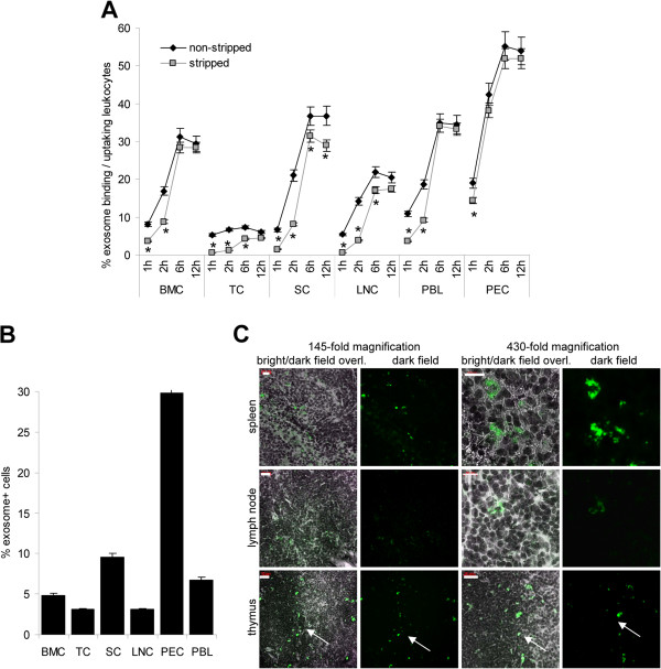
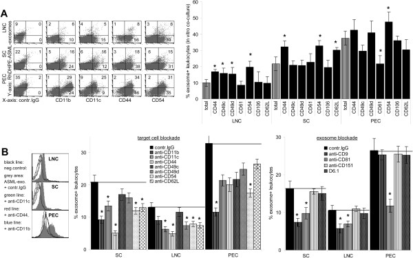
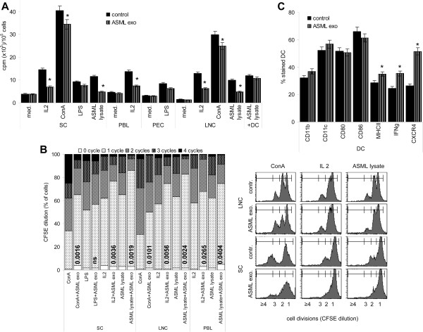
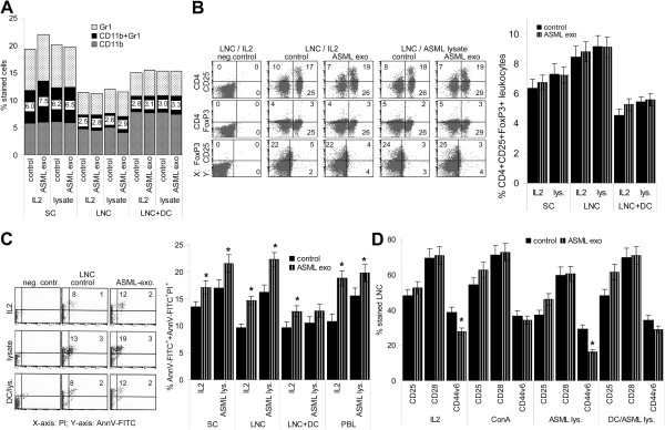
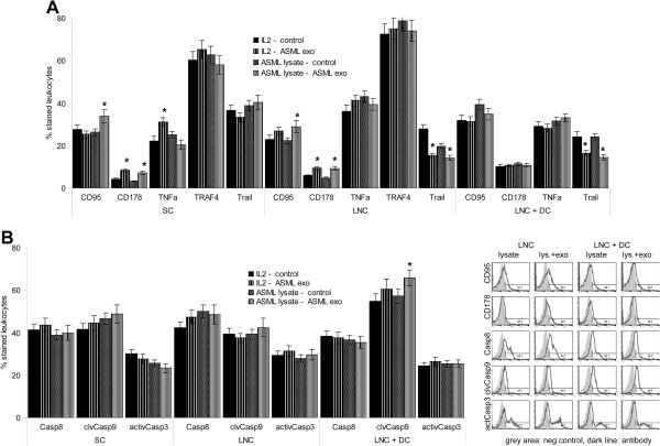
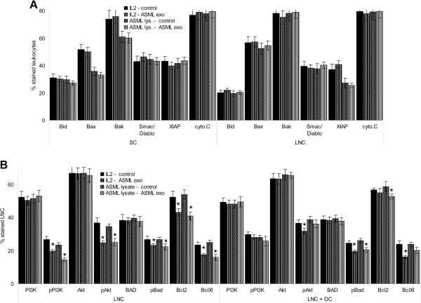
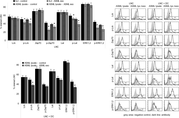
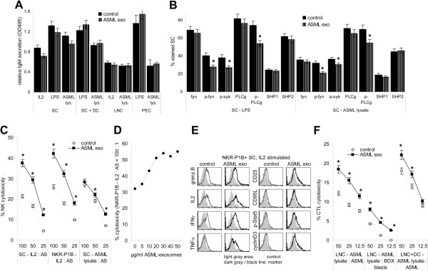
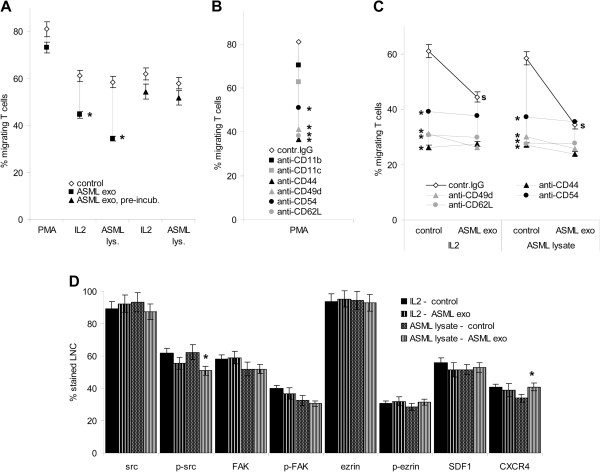
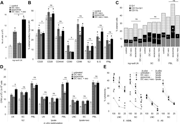
Results: ASML-exosomes bind to and are taken up by all leukocyte subpopulations in vivo and in vitro, uptake by CD11b+ leukocytes exceeding that by T and B cells. ASML-exosomes affect leukocyte proliferation via reduced CD44v6 up-regulation and lck, ZAP70 and ERK1,2 phosphorylation, which can be compensated by dendritic cells (DC). ASML-exosomes do not support Treg. Yet, impaired activation of anti-apoptotic signals is accompanied by slightly increased apoptosis susceptibility. IgM secretion is unaffected; NK and CTL activity are strengthened, ASML-exosomes co-operating with DC in CTL activation. ASML-exosomes transiently interfere with leukocyte migration by occupying migration-promoting receptors CD44, CD49d, CD62L and CD54 during binding/internalization.

Conclusion: ASML-exosomes might well serve as adjuvant in immunotherapy as they support leukocyte effector functions and have only a minor impact on leukocyte activation, which can be overridden by DC. However, exosome-induced modulation of immune cells relies, at least in part, on exosome uptake and message transfer. This implies that depending on the individual tumor's exosome composition, exosomes may distinctly affect the immune system. Nonetheless, whether immunotherapy can profit from using tumor-exosomes as adjuvant can easily be settled beforehand in vitro.


Figures

Figure 1
Figure 1
Tumor-exosome uptake by leukocytes. (A) BMC, TC, SC, LNC, PBL and PEC were incubated at 37°C with RhDHPE-labeled ASML-exosomes for the indicated periods. Exosome binding and uptake (fluorescence after 2 acid washes) was evaluated by flow-cytometry: Mean percent ± SD (3 experiments) of exosome+ leukocytes. Significant differences between exosome binding/uptake versus uptake: *. (B,C) SP-Dio18(3)-labeled exosomes (200 μg) were injected i.v. and hematopoietic organs were excised after 24 h: (C) Mean percent ± SD (3 rats) of exosome+ leukocytes evaluated by flow-cytometry. (D) Confocal microscopy showing representative examples of exosome+ cells in shock frozen lymph node, spleen and thymus (arrow: boundary cortex/medulla) sections (scale bar left: 20 μm, right: 10 μm). Tumor-exosomes bind and are taken up in vitro and in vivo by cells from all hematopoietic organs. Uptake is most rapid and abundant in PEC.
Figure 2
Figure 2
Tumor-exosome uptake by leukocyte subpopulations. (A) LNC, SC, PBL and PEC were incubated with RhDHPE-labeled ASML-exosomes for 6 h and stained with leukocyte subset-specific antibodies. The mean percent ± SD of marker+exosome+ / marker+ cells (3 experiments) is shown. (B,C) In vivo uptake of SP-Dio18(3) labeled ASML-exosomes 24 h after i.v. application: (B) flow-cytometry of dispersed cells. The mean percent ± SD of marker+exosome+ / marker+ cells (3 rats) is shown. (C) Confocal microscopy of shock frozen tissue sections (spleen sections were stained with anti-sIgM, lymph node sections with anti-CD4, -CD8, -CD11b and -CD11c. For lymph node sections stained with anti-CD4 and anti-CD11b a higher magnification is included (scale bar spleen: 20 μm, lymph nodes: 10 μm). (A,B) Significant differences in the % exosome+marker+ cells as compared to exosome+ cells in the total organ: *. Exosomes are taken up by cells of all major leukocyte subpopulations.
Figure 3
Figure 3
Adhesion molecules engaged in tumor-exosome uptake. (A) Cells as in (Figure 2A) were stained with adhesion molecule-specific antibodies: representative examples and mean percent ± SD of marker+exosome+ / marker+ cells (3 experiments). (B) LNC, SC and PEC or exosomes were pre-incubated with the indicated antibodies (30 min, 4°C). After washing, cells were co-incubated with dye-labeled exosomes 2h, 4°C: representative examples and mean percent ± SD (3 experiments) of exosome+ cells. (A) Significant differences in the % exosome+marker+ cells as compared to exosome+ cells in the total organ: *, (B) significant differences compared to control IgG treatment: *. There is evidence for an engagement of CD11b, CD11c, CD44, CD49d, CD54 and CD62L in exosome uptake.
Figure 4
Figure 4
ASML-exosomes and leukocyte proliferation. Lymphocytes were stimulated for 72 h as indicated with/without ASML-exosomes. Where indicated, cultures additionally contained ASML lysate-pulsed DC (LNC:DC = 10:1). (A) Mean ± SD (triplicates) of 3H-thymidine incorporation. (B) Examples of CFSE dilution in LNC and SC cultured for 72 h and mean percentage (triplicates) of cells that did progress through 0–4 cycles. Significant differences to cultures not containing ASML-exosomes are shown. (C) BMC-derived DC were cultured as described in MM. During the last 24 h of culture in the presence of LPS, ASML-exosomes were added where indicated: Mean percent ± SD (3 experiments) of CD11b+, CD11c+, CD80+, CD86+, MHCII+, IFNγ+ and CXCR4+ cells (flow-cytometry). (A,C) Significant differences in the presence of ASML-exosomes: *. Exosomes inhibit lymphocyte proliferation, which can be circumvented by activated DC.
Figure 5
Figure 5
ASML-exosomes, immunosuppression, apoptosis and activation markers. Leukocytes were stimulated as described in Figure 4. (A) Mean percent (3 experiments) of Gr1+, CD11b+ and Gr1+CD11b+ (MDSC) cells. (B) examples of CD4+CD25+, CD4+FoxP3+ and CD25+FoxP3+ cells and mean percent ± SD (3 experiments) of CD4+CD25+FoxP3+ cells. (C) Representative examples of AnnexinV/PI staining and mean percent ± SD (3 experiments) of AnnV-FITC+/AnnV-FITC+/PI+ cells. (D) Mean percent ± SD (3 experiments) of CD25+, CD28+ and CD44v6+ cells. (C,D) Significant differences in the presence of ASML-exosomes: *. There is no evidence for ASML-exosomes affecting MDSC or Treg. However, apoptosis susceptibility is slightly increased and expansion of CD44v6+ cells is impaired.
Figure 6
Figure 6
ASML-exosomes and death receptors. Leukocytes were stimulated as described in Figure 4. Expression of (A) death receptors and (B) caspases was evaluated by flow-cytometry. Representative examples and mean percent ± SD (3 experiments) of stained cells; significant differences in the presence of ASML-exosomes: *. ASML-exosomes do not promote caspase activation.
Figure 7
Figure 7
ASML-exosomes and activation of pro- and anti-apoptotic molecules. Leukocytes were stimulated as described in Figure 4. Expression of (A) pro-apoptotic molecules and (B) anti-apoptotic molecules was evaluated by flow-cytometry. Mean percent ± SD (3 experiments) of stained cells; significant differences in the presence of ASML-exosomes: *. ASML-exosomes slightly affect activation of the PI3K/Akt pathway.
Figure 8
Figure 8
ASML-exosomes and TCR activation. Leukocytes were stimulated as described in Figure 4. Expression of molecules involved in TCR signaling was evaluated by flow-cytometry. Representative examples and mean percent ± SD (3 experiments) of stained cells; significant differences in the presence of ASML-exosomes: *. In the presence of ASML-exosomes activation of lck and TCR downstream kinases of the MAPK pathway is impaired. Both effects are strongly mitigated in the presence of DC.
Figure 9
Figure 9
ASML-exosomes and effector lymphocytes. (A,B) SC, LNC and PEC were stimulated with IL2, LPS or ASML lysate with/without ASML-exosomes: (A) Supernatants were harvested after 4d to evaluate IgM secretion by ELISA. (B) Expression of B cell activation-related signal transduction molecules was evaluated by flow-cytometry after 2d (mean values ± SD, 3 experiments). (C) SC and NKR-P1B+ cells were cultured in the presence of 100U IL2/ml or ASML-lysate for 2d. NK cytotoxicity was evaluated with 3H-thymidine labeled AS target cells. (D) NKR-P1B+ cells were cultured in the presence of 100U IL2/ml and titrated amounts of ASML-exosomes. Cytotoxicity was evaluated as in (C) after 2d of co-culture. (E) NKR-P1B+ cells were cultured in the presence of 100U IL2/ml and 40 μg/ml ASML-exosomes for 2d. Expression of granzymeB, IL2, IFNγ, TNFα, CD25, CD95L, p-Stat5 and CyclinD3 was evaluated by flow cytometry. (F) LNC were cultured in the presence of ASML-lysate for 8d. Where indicated, cultures contained ASML lysate-loaded DC. CTL activity was evaluated with 3H-thymidine labeled ASML and BDX blast target cells: (C,D,F) Mean percent ± SD (triplicates) of cytotoxicity at the indicated E:T ratios. (A-F) Significant differences in cultures containing ASML-exosomes: *. Tumor-exosomes do not hamper a primary B cells response to T cell-dependent or T cell-independent stimuli. But, activation of fyn, syk and PLCγ are slightly impaired. Tumor-exosomes strengthen NK and CTL activity.
Figure 10
Figure 10
ASML-exosomes and T cell migration. T cells were stimulated as indicated for 24 h. (A-C) Stimulated T cells were seeded in RPMI/1%FCS in the upper part of a Boyden chamber, the lower part contained RPMI/20%FCS. Where indicated, T cells stimulated in the presence of ASML-exosomes were washed, evaluating migration in the absence of ASML-exosomes. The percentage of migrating cells was evaluated after 4 h at 37°C. (B) T cells were stimulated for 24 h with PMA. Before migration, cells were incubated with the indicated antibodies (30 min, 4°C). (C) T cells were stimulated, washed and incubated with the indicated antibodies (30 min, 4°C). Migration was evaluated in the presence/absence of ASML-exosomes. (A-C) Mean percent ± SD (triplicates, 3 experiments) of migrating cells. (A) Significant differences in cultures containing or pre-incubated with ASML-exosomes: *, (B,C) significant antibody inhibition: *, (C) significant inhibition in the presence of ASML-exosomes: s. (D) Migration-related signaling molecule including SDF1 and CXCR4 expression was evaluated by flow-cytometry. Mean percent ± SD (3 experiments) of stained cells, significant differences in cultures containing ASML-exosomes: *. Tumor-exosomes affect T cell migration. This is due to a blockade of migration-relevant adhesion molecules engaged in exosome uptake. Up-taken exosomes hardly affect T cell migration.
Figure 11
Figure 11
In vivo impact of ASML-exosomes on leukocyte activation. Rats received 3-times 2x10⁶ DC, subcutaneously and/or 7-times 500 μg ASML-exosomes, i.v. as described in MM. Rats were sacrificed 3d after the 3rd DC application to analyze the draining LNC, SC and PBL. (A) Number of draining LNC (mean ± SD, 3 rats), (B) leukocyte activation markers, IL2, IL12 and IFNγ expression (flow cytometry, mean ± SD, 3 rats and representative examples), (C) MDSC (flow cytometry, mean ± SD, 3 rats), (D) 3H-thymidine incorporation after 3d in vitro culture and (E) cytotoxic activity against ASML and AS (NK susceptible) targets after 10d in vitro culture in the presence of ASML lysate (mean ± SD, triplicates). (A-E) Significant differences to lymphocytes from untreated rats: *, differences between lymphocytes from rats receiving DC or DC plus ASML-exosomes are indicated as ns (not significant) or s (significant, p <0.01). In vivo, ASML-exosomes support recruitment and/or proliferation of draining LNC and CD11b, CD86, IL12 and IFNγ expression. Despite an increase in MDSC in the peripheral blood, ASML-exosomes support DC vaccination-induced T cell expansion and cytotoxic activity.

References

    1. Mathivanan S, Ji H, Simpson RJ. Exosomes: extracellular organelles important in intercellular communication. J Proteomics. 2010;73:1907–1920. doi: 10.1016/j.jprot.2010.06.006. - DOI - PubMed
    1. Cocucci E, Racchetti G, Meldolesi J. Shedding microvesicles: artefacts no more. Trends Cell Biol. 2009;19:43–51. doi: 10.1016/j.tcb.2008.11.003. - DOI - PubMed
    1. Pap E, Pállinger E, Pásztói M, Falus A. Highlights of a new type of intercellular communication: microvesicle-based information transfer. Inflamm Res. 2009;58:1–8. - PubMed
    1. Ahmed KA, Xiang J. Mechanisms of cellular communication through intercellular protein transfer. J Cell Mol Med. 2011;15:1458–1473. doi: 10.1111/j.1582-4934.2010.01008.x. - DOI - PMC - PubMed
    1. Seow Y, Wood MJ. Biological gene delivery vehicles: beyond viral vectors. Mol Ther. 2009;17:767–777. doi: 10.1038/mt.2009.41. - DOI - PMC - PubMed