. 2013 May;133(5):1269-77.
doi: 10.1038/jid.2012.421. Epub 2012 Nov 29.

Oncogenic B-RAF(V600E) signaling induces the T-Box3 transcriptional repressor to repress E-cadherin and enhance melanoma cell invasion

Affiliations

Oncogenic B-RAF(V600E) signaling induces the T-Box3 transcriptional repressor to repress E-cadherin and enhance melanoma cell invasion

Suzanah C Boyd et al. J Invest Dermatol. 2013 May.

Abstract

Approximately 50% of melanomas require oncogenic B-RAF(V600E) signaling for proliferation, survival, and metastasis, and the use of highly selective B-RAF inhibitors has yielded remarkable, although short-term, clinical responses. Reactivation of signaling downstream of B-RAF is frequently associated with acquired resistance to B-RAF inhibitors, and the identification of B-RAF targets may therefore provide new strategies for managing melanoma. In this report, we applied whole-genome expression analyses to reveal that oncogenic B-RAF(V600E) regulates genes associated with epithelial-mesenchymal transition in normal cutaneous human melanocytes. Most prominent was the B-RAF-mediated transcriptional repression of E-cadherin, a keratinocyte-melanoma adhesion molecule whose loss is intimately associated with melanoma invasion and metastasis. Here we identify a link between oncogenic B-RAF, the transcriptional repressor Tbx3, and E-cadherin. We show that B-RAF(V600E) induces the expression of Tbx3, which potently represses E-cadherin expression in melanocytes and melanoma cells. Tbx3 expression is normally restricted to developmental embryonic tissues and promoting cell motility, but it is also aberrantly increased in various cancers and has been linked to tumor cell invasion and metastasis. We propose that this B-RAF/Tbx3/E-cadherin pathway has a critical role in promoting the metastasis of B-RAF-mutant melanomas.


Figures

Figure 1
Figure 1. Activity of exogenous B-RAFV600E in human melanocytes
Primary human melanocytes were infected with lentiviruses encoding B-RAFV600E with copGFP or copGFP alone (control) for three days. Expression levels of phosphorylated ERK (p-ERK) were compared to those observed in a series of melanoma cell lines with known B-RAF/N-RAS genotypes (WMM1175: N-RASG13R; NM39, SKMel28 and ME1042: B-RAFV600E). Ectopic B-RAF expression was detected by probing for the Myc-tag of B-RAFV600E.
Figure 2
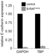
Figure 2. B-RAFV600E signaling represses E-cadherin
(a) Melanocytes were infected with lentiviruses encoding B-RAFV600E with copGFP or copGFP alone (control) for three days. Total RNA was analysed by qRT-PCR. E-cadherin transcript levels in B-RAFV600E-transduced melanocytes were normalised to levels of GAPDH or TBP housekeeping genes and expressed relative to normalized E-cadherin transcript levels in control-transduced cells. (b) Three independent melanocyte strains and NM179 melanoma cells were infected with lentiviruses encoding B-RAFV600E with copGFP or copGFP alone (control) for three days. Total protein was immunoblotted for the indicated proteins. (c) B-RAF signaling was inhibited in the B-RAF mutant melanoma cell lines, NM176 and ME1042, using the B-RAFV600E-specific silencing molecule for five days or 10μM PLX4032 for two days. Total protein was immunoblotted for the indicated proteins.
Figure 3
Figure 3. B-RAFV600E regulates Tbx3 expression in melanocytes
Primary human melanocytes were infected with lentiviruses encoding B-RAFV600E with copGFP or copGFP alone (control) for three days. (a) Transcript expression levels of established E-cadherin transcriptional regulators in B-RAFV600E-transduced melanocytes measured in gene expression arrays. Transcript levels are expressed relative to control transduced melanocytes (indicated by grey line). (b) Total RNA derived from a minimum of three independent transduction experiments were analysed by qRT-PCR. Tbx3 transcript levels in B-RAFV600E-transduced melanocytes were normalised to levels of GAPDH or TBP housekeeping genes and expressed relative to normalised Tbx3 transcript levels in control-transduced cells. (c) Tbx3 cDNA derived from B-RAFV600E or control-transduced melanocytes was amplified for 25 PCR cycles and products analysed using agarose gel electrophoresis.
Figure 4
Figure 4. B-RAFV600E regulates Tbx3 expression in melanoma
(a) 501mel melanoma cells were transfected with the human Tbx3 promoter (−249 to +168 in pGL3basic) or the pGL3 basic vector alone together with 25 or 100 ng of B-RAF wild type or V600E expression plasmid. Promoter activity was derived form the measured luciferase activity normalized to the promoter activity of pGL3 basic transfected cells. Immunoblotting confirmed similar expression of the Myc epitope-tagged B-RAF constructs using anti-Myc antibody. (b) Total RNA from stage III melanoma lymph node metastasis was analysed by gene expression arrays. The relative median Tbx3 transcript expression levels are shown (n=27 B-RAF wild type (wt) tumors and n=33 tumors expressing B-RAFV600E). For comparison expression levels of Tbx3 in cultured normal melanocytes transduced to express control or B-RAFV600E are presented (black asterisk).
Figure 5
Figure 5. B-RAF mediates regulation of E-cadherin via Tbx3
Melanocytes were transduced with lentiviruses containing the indicated shRNA constructs. Five days post infection the cells were re-transduced with copGFP control lentivirus or lentiviruses expressing B-RAFV600E or copGFP, as shown. Total protein was immunoblotted for the indicated proteins. This figure is compiled from duplicate immunoblots.
Figure 6
Figure 6. B-RAFV600E promotes melanoma cell invasion
The V600E B-RAF mutant allele or Tbx3 were specifically silenced in the NM176 and ME1042 melanoma cell lines for five days. The invasion of melanoma cells was determined by transwell matrigel invasion assays and is presented as relative invasion compared to control silenced melanoma cells. Knockdown of BRAFV600E and Tbx3 induced a significant (**p<0.05) or near significant (*p=0.05) decrease in cell invasion (p-values: NM176 V600E: 0.003, Tbx3: 0.0007; ME1042 V600E: 0.04, Tbx3: 0.05).

Comment in

References

    1. Balch CM, Soong SJ, Gershenwald JE, Thompson JF, Reintgen DS, Cascinelli N, et al. Prognostic factors analysis of 17,600 melanoma patients: validation of the american joint committee on cancer melanoma staging system. J Clin Oncol. 2001;19:3622–3634. - PubMed
    1. Bamshad M, Lin RC, Law DJ, Watkins WC, Krakowiak PA, Moore ME, et al. Mutations in human TBX3 alter limb, apocrine and genital development in ulnar-mammary syndrome. Nat Genet. 1997;16:311–315. - PubMed
    1. Basolo F, Torregrossa L, Giannini R, Miccoli M, Lupi C, Sensi E, et al. Correlation between the BRAF V600E mutation and tumor invasiveness in papillary thyroid carcinomas smaller than 20 millimeters: analysis of 1060 cases. J Clin Endocrinol Metab. 2010;95:4197–4205. - PubMed
    1. Becker TM, Philipsz S, Scurr LL, Fung C, Haferkamp S, Kefford RF, et al. Oncogenic B-RAF(V600E) promotes anchorage-independent survival of human melanocytes. J Invest Dermatol. 2010;130:2144–2147. - PubMed
    1. Bolos V, Peinado H, Perez-Moreno MA, Fraga MF, Esteller M, Cano A. The transcription factor Slug represses E-cadherin expression and induces epithelial to mesenchymal transitions: a comparison with Snail and E47 repressors. J Cell Sci. 2003;116:499–511. - PubMed

Publication types

MeSH terms