

doi: 10.1107/S1744309112041759.
Epub 2012 Nov 14.
Interactions of Mn2+ with a non-self-complementary Z-type DNA duplex
Affiliations
- PMID: 23192018
- PMCID: PMC3509959
- DOI: 10.1107/S1744309112041759
Item in Clipboard
Interactions of Mn2+ with a non-self-complementary Z-type DNA duplex
Acta Crystallogr Sect F Struct Biol Cryst Commun.
.
Abstract
Crystal structures of the hexanucleotide d(CACGCG)·d(CGCGTG) were determined in two crystal lattices when different concentrations of the counterion Mn2+ were used in crystallization. The availability of Mn2+ during the crystallization process appears to play an important role in inducing different crystal packings that lead to crystals belonging to the two space groups P2(1) and P6(5). Analysis of the molecular interactions of Mn2+ with the Z-form duplexes shows direct coordination to the purine residues G and A.
Figures
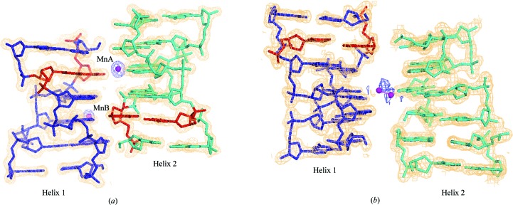
(a) Helix 1 (blue), helix 2 (cyan) and two Mn2+ ions in Mn_P21. (b) Helix 1 (blue), an arbitrary helix 2 (cyan) comprising dinucleotide steps (x, y, z), (y, −x + y, z + 1/6) and (−x + y, −x, z + 1/3) and a disordered Mn2+ ion in Mn_P65. The anomalous difference Fourier map (blue) and 2F
o − F
c map (orange; for the helices only) are contoured at 3σ and 1σ levels, respectively. The A2·T11 base pairs are coloured red. The Mn2+ ions are shown as spheres. The water molecules are omitted for clarity.

Packing of the helical columns in Mn_P21 (a, b) and Mn_P65 (c, d). (a) and (c) are views down the helical axis, while (b) and (d) are views perpendicular to the helical axis. Helix 1 and helix 2 [except in (a)] are coloured blue and cyan, respectively. The A·T base pairs in the ordered hexamer helices are coloured red. Spheres represent Mn2+ bound to helices. The column with the 65 screw symbol indicates the disordered column built from the dinucleotide step. No direction for these columns could be assigned since the position of the A·T base pair is disordered. The unit cell is shown as thin lines.

Interactions of ions: (a) MnA and (b) MnB in Mn_P21 and (c) the disordered Mn2+ ion in Mn_P65 (viewed down the helical axis). The Mn2+ ions are shown as spheres, water molecules are shown as crosses, ion-mediated and water-mediated interactions are shown as red dashed lines and hydrogen bonds of base pairs are shown as black dashed lines. The ion induces distortion of the hydrogen-bond base-pair schemes in (b) and (c).
References
-
- Anderson, J. A., Kuntz, G. P., Evans, H. H. & Swift, T. J. (1971). Biochemistry, 10, 4368–4374. - PubMed
-
- Bartels, K. S. & Klein, C. (2003). The Automar Manual v.1.4. Norderstedt, Germany: MAR Research GmbH.
Publication types
MeSH terms
Substances
Associated data
- Actions
- Actions
LinkOut - more resources
Full Text Sources
Research Materials
