

doi: 10.1107/S1744309112045563.
Epub 2012 Nov 28.
Production, purification, crystallization and structure determination of H-1 Parvovirus
Affiliations
- PMID: 23192051
- PMCID: PMC3509992
- DOI: 10.1107/S1744309112045563
Item in Clipboard
Production, purification, crystallization and structure determination of H-1 Parvovirus
Acta Crystallogr Sect F Struct Biol Cryst Commun.
.
Abstract
Crystals of H-1 Parvovirus (H-1PV), an antitumor gene-delivery vector, were obtained for DNA-containing capsids and diffracted X-rays to 2.7 Å resolution using synchrotron radiation. The crystals belonged to the monoclinic space group P2(1), with unit-cell parameters a=255.4, b=350.4, c=271.6 Å, β=90.34°. The unit cell contained two capsids, with one capsid per crystallographic asymmetric unit. The H-1PV structure has been determined by molecular replacement and is currently being refined.
Figures
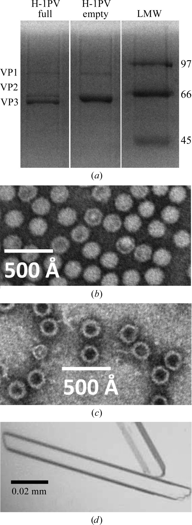
Purification and crystallization of H-1PV full and empty capsids. (a) A 15% SDS–PAGE gel of the purified H-1PV capsids showing the relative ratios and positions of VP1, VP2 and VP3 (molecular weights of 81, 65 and 63 kDa, respectively). The positions of low-molecular-weight standards (labeled in kDa; Bio-Rad, Hercules, California, USA) are indicated on the right-hand side. (b, c) Negatively stained electron micrographs of H-1PV full capsids viewed at 100 000× magnification (b) and H-1PV empty capsids viewed at 60 000× magnification (c). (d) Optical photograph of H-1PV full capsid crystals in a rod-shaped habit.
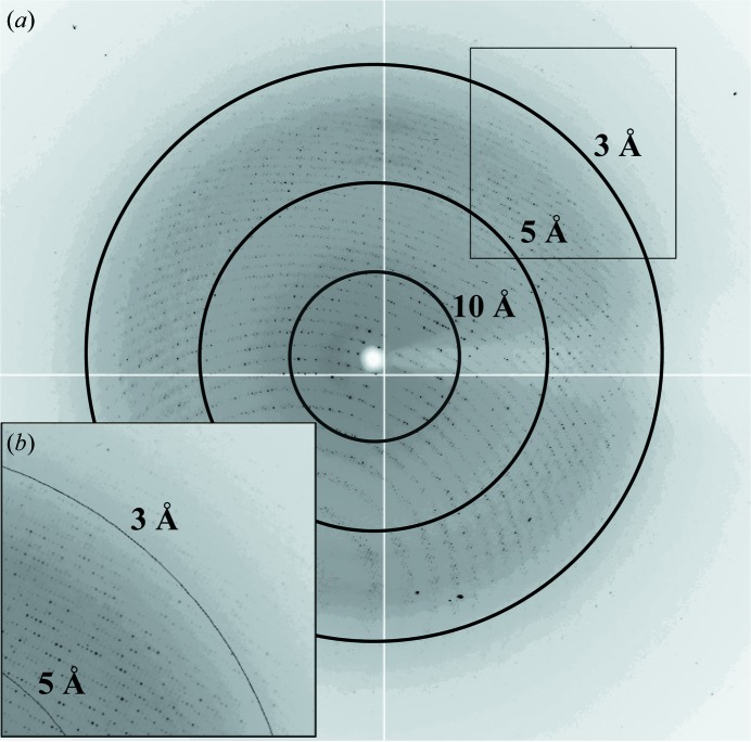
X-ray diffraction image for a crystal of H-1PV full capsids. The image is a typical 0.3° oscillation photograph. The concentric rings indicate the 10.0, 5.0 and 3.0 Å resolution shells. (b) The inset shows a close-up of the boxed region in the upper right-hand corner.
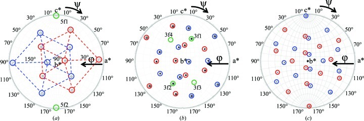
Stereographic projections of the self-rotation function search for the H-1PV full capsid X-ray diffraction data. (a) κ = 72°, (b) κ = 120° and (c) κ = 180°, searching for fivefold, threefold and twofold icosahedral symmetry elements, respectively. The peaks representing fivefold positions are delineated by the dashed pentagons in (a). The peaks belonging to each of the two viral capsids in the unit cell are circled in red and blue, respectively. The peaks interpreted as Klug peaks (5f1, 5f2 and 3f1–4) are circled in green and labeled in (a) and (b). The a*, b* and c* axes are labeled.
Similar articles
-
Structural characterization of H-1 parvovirus: comparison of infectious virions to empty capsids.J Virol. 2013 May;87(9):5128-40. doi: 10.1128/JVI.03416-12. Epub 2013 Feb 28. J Virol. 2013. PMID: 23449783 Free PMC article.
-
Production, purification and preliminary X-ray crystallographic studies of adeno-associated virus serotype 9.Acta Crystallogr Sect F Struct Biol Cryst Commun. 2009 Jul 1;65(Pt 7):715-8. doi: 10.1107/S1744309109021460. Epub 2009 Jun 27. Acta Crystallogr Sect F Struct Biol Cryst Commun. 2009. PMID: 19574648 Free PMC article.
-
Production, purification, crystallization and preliminary X-ray analysis of adeno-associated virus serotype 8.Acta Crystallogr Sect F Struct Biol Cryst Commun. 2005 Jun 1;61(Pt 6):558-61. doi: 10.1107/S1744309105014132. Epub 2005 Jun 1. Acta Crystallogr Sect F Struct Biol Cryst Commun. 2005. PMID: 16511095 Free PMC article.
-
Production, purification and preliminary X-ray crystallographic studies of adeno-associated virus serotype 7.Acta Crystallogr Sect F Struct Biol Cryst Commun. 2007 Dec 1;63(Pt 12):1073-6. doi: 10.1107/S1744309107060289. Epub 2007 Nov 30. Acta Crystallogr Sect F Struct Biol Cryst Commun. 2007. PMID: 18084098 Free PMC article.
-
Crystallization of RNA.Cell Mol Life Sci. 2001 Feb;58(2):234-43. doi: 10.1007/PL00000851. Cell Mol Life Sci. 2001. PMID: 11289305 Free PMC article. Review.
Cited by
-
Structural characterization of H-1 parvovirus: comparison of infectious virions to empty capsids.J Virol. 2013 May;87(9):5128-40. doi: 10.1128/JVI.03416-12. Epub 2013 Feb 28. J Virol. 2013. PMID: 23449783 Free PMC article.
-
A novel scalable, robust downstream process for oncolytic rat parvovirus: isoelectric point-based elimination of empty particles.Appl Microbiol Biotechnol. 2017 Apr;101(8):3143-3152. doi: 10.1007/s00253-016-8071-x. Epub 2017 Jan 14. Appl Microbiol Biotechnol. 2017. PMID: 28091791 Free PMC article.
-
Structure of neurotropic adeno-associated virus AAVrh.8.J Struct Biol. 2015 Oct;192(1):21-36. doi: 10.1016/j.jsb.2015.08.017. Epub 2015 Aug 31. J Struct Biol. 2015. PMID: 26334681 Free PMC article.
-
Parvoviruses cause nuclear envelope breakdown by activating key enzymes of mitosis.PLoS Pathog. 2013 Oct;9(10):e1003671. doi: 10.1371/journal.ppat.1003671. Epub 2013 Oct 31. PLoS Pathog. 2013. PMID: 24204256 Free PMC article.
-
Stability and safety key factors of the oncolytic protoparvovirus H-1 from manufacturing to human application.Appl Microbiol Biotechnol. 2023 Aug;107(15):4777-4787. doi: 10.1007/s00253-023-12521-4. Epub 2023 May 20. Appl Microbiol Biotechnol. 2023. PMID: 37209160 Free PMC article.
References
-
- Agbandje, M., McKenna, R., Rossmann, M. G., Strassheim, M. L. & Parrish, C. R. (1993). Proteins, 16, 155–171. - PubMed
-
- Agbandje-McKenna, M. & Chapman, M. S. (2006). Parvoviruses, edited by J. R. Kerr, S. F. Cotmore, M. E. Bloom, R. M. Linden & C. R. Parrish, pp. 125–139. London: Hodder Arnold.
-
- Agbandje-McKenna, M., Llamas-Saiz, A. L., Wang, F., Tattersall, P. & Rossmann, M. G. (1998). Structure, 6, 1369–1381. - PubMed
-
- Angelova, A. L., Aprahamian, M., Grekova, S. P., Hajri, A., Leuchs, B., Giese, N. A., Dinsart, C., Herrmann, A., Balboni, G., Rommelaere, J. & Raykov, Z. (2009). Clin. Cancer Res. 15, 511–519. - PubMed
Publication types
MeSH terms
Substances
Grants and funding
LinkOut - more resources
Full Text Sources
Other Literature Sources
