Review
. 2013 May 21;42(10):4310-26.
doi: 10.1039/c2cs35401b. Epub 2012 Nov 28.

Carbohydrate microarrays

Affiliations
Review

Carbohydrate microarrays

Sungjin Park et al. Chem Soc Rev. .

Abstract

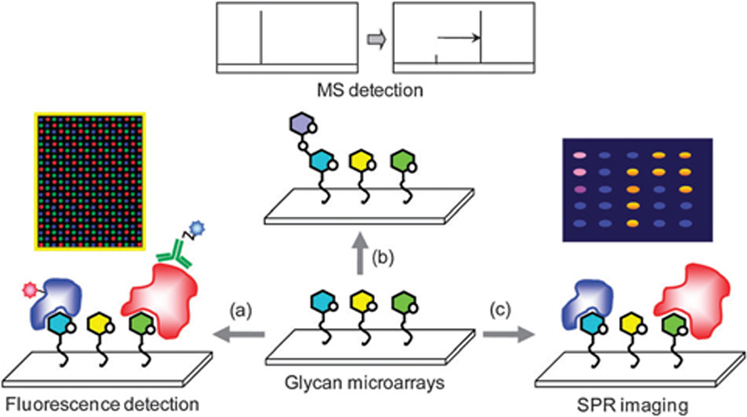
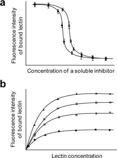
In the last decade, carbohydrate microarrays have been core technologies for analyzing carbohydrate-mediated recognition events in a high-throughput fashion. A number of methods have been exploited for immobilizing glycans on the solid surface in a microarray format. This microarray-based technology has been widely employed for rapid analysis of the glycan binding properties of lectins and antibodies, the quantitative measurements of glycan-protein interactions, detection of cells and pathogens, identification of disease-related anti-glycan antibodies for diagnosis, and fast assessment of substrate specificities of glycosyltransferases. This review covers the construction of carbohydrate microarrays, detection methods of carbohydrate microarrays and their applications in biological and biomedical research.


Figures

Fig. 1
Fig. 1
Glycoconjugates in cells.
Fig. 2
Fig. 2
Functionalization of glycans via glycosidic linkages (I), N-hydroxylamines(II) and reductive amination (III).
Fig. 3
Fig. 3
Immobilization strategy for construction of glycan microarrays. (a) Noncovalent, site-nonspecific attachment of glycans on the surface, (b) noncovalent, site-specific attachment of glycans on the surface, (c) covalent, sitenonspecific attachment of glycans on the surface, and (d) covalent, site-specific attachment of glycans on the surface.
Fig. 4
Fig. 4
Noncovalent and site-nonspecific immobilization. (a) Attachment of unmodified polysaccharides to the surface, (b) attachment of free heparin polysaccharides to the poly-L-lysine-coated surface, and (c) attachment of modified dextrans to the amine or semicarbazide-coated surface.
Fig. 5
Fig. 5
Noncovalent and site-specific immobilization. (a) Attachment of lipid-conjugated glycans to nitrocellulose, (b) attachment of fluorous-tagged sugars to the fluoroalkylated surface, (c) attachment of fluorous-tagged glycans to fluorous phosphonate derivatized on the aluminum oxide-coated surface, (d) attachment of biotin-linked sugars to the streptavidin-coated surface, and (e) attachment of oligonucleotide-conjugated glycans to the complementary oligonucleotide-coated surface.
Fig. 6
Fig. 6
Covalent and site-nonspecific immobilization. Attachment of free glycans on (a) aryltrifluoromethyldiazirine, (b) 4-azido-2,3,5,6-tetrafluorophenyl group, (c) phthalimide-derivatized surfaces by UV irradiation, and (d) phenylboronic acid-coated surfaces.
Fig. 7
Fig. 7
Covalent and site-specific immobilization. (a) Attachment of maleimide-conjugated glycans to the thiol-coated surface, (b) attachment of thiol-linked glycans to the maleimide-coated surface, (c) attachment of thiosulfonate-conjugated glycans to thiol-coated surface, (d) attachment of thiol-conjugated sugars to the 2-pyridyl disulfide-coated surface, (e) attachment of cyclopentadiene-linked sugars to the benzoquinone-coated surface, (f) attachment of dienophile-conjugated sugars to the tetrazine-coated surface, (g) attachment of p-aminophenyl group-linked sugars to the cyanuric chloride-coated surface, (h) attachment of amine-linked sugars to the NHS ester-coated surface, (i) attachment of aminooxy-linked chondroitin oligosaccharides to the aldehyde-coated surface, and (j) attachment of heparin oligosaccharides, which are obtained from nitrous acid depolymerization of heparin, to the amine-coated surface.
Fig. 8
Fig. 8
Covalent and site-specific immobilization. (a) Attachment of hydrazide-linked sugars to the epoxide-coated surface, (b) attachment of glycan-linked BSA to the epoxide-coated surface, (c) attachment of azide-linked glycans to the alkyne-coated surface, (d) attachment of alkyne-linked glycans to the azidecoated surface, (e) attachment of azide-linked glycans to the phosphane-coated surface via Staudinger reaction, and (f) attachment of 4-azido-2,3,5,6tetrafluorophenyl group-conjugated glycans to the polymer monolayer by photochemistry.
Fig. 9
Fig. 9
(a) Immobilization of glycans obtained from one-step reactions on the NHS ester-derivatized surface and (b) immobilization of unmodified glycans on the hydrazide or aminooxy-derivatized surface.
Fig. 10
Fig. 10
Glycan presentation on the solid surface. Multivalent binding is critical for strong glycan–protein recognition events. To form a high avidity multivalent complex, the spacing and orientation of glycans on the surface must match the spacing and orientation of binding sites on a GBP. (a) A lectin with short spacing between binding sites may bind strongly to the high density of glycans on the surface. (b) A lectin with short spacing between binding sites may not bind strongly to the low density of glycans on the surface. However, an antibody with longer spacing between binding sites can bind well to glycans at either high or low density. (c) Glycan microarrays can be constructed by immobilizing multivalent glycoconjugates such as glycodendrimers, neoglycoproteins/neoglycopeptides and glycopolymers. Use of multivalent glycoconjugates provides unique opportunities to modulate glycan presentation.
Fig. 11
Fig. 11
Detection methods of glycan microarrays. (a) Binding events of fluorophore-labeled proteins to glycans on microarrays can be monitored by using a fluorescence scanner. (b) Enzymatic reactions on microarrays can be detected using MS. (c) SPR imaging can be used as a label-free detection method for glycan microarrays.
Fig. 12
Fig. 12
Application of glycan microarrays for rapid analysis of glycan–protein interactions.
Fig. 13
Fig. 13
Application of glycan microarrays for detection of viruses and whole cells.
Fig. 14
Fig. 14
Determination of (a) IC50 values of soluble inhibitors and (b) apparent dissociation constants (Kd values) for glycan–protein interactions using glycan microarrays.
Fig. 15
Fig. 15
Glycan binding properties of human intravenous immunoglobulin G (IgG). The antibody binds to a variety of glycans on the microarrays.
Fig. 16
Fig. 16
Detection of aberrant glycans displayed on proteins.

References

    1. Varki A, Glycobiology, 1993, 3, 97–130. - PMC - PubMed
    1. Bertozzi CR and Kiessling LL, Science, 2001, 291, 2357–2364. - PubMed
    1. Park S, Lee M-R and Shin I, Chem. Soc. Rev, 2008, 37, 1579–1591. - PubMed
    1. Essentials of Glycobiology, ed. Varki A, Cummings R, Esko J, Freeze H, Stanley P, Bertozzi CR, Hart G and Etzler ME, Cold Spring Harbor Laboratory Press, 2nd edn, 2009. - PubMed
    1. Crocker PR, Curr. Opin. Pharmacol, 2005, 5, 431–437. - PubMed

Publication types

LinkOut - more resources