Review
. 2013 Feb;224(3):335-58.
doi: 10.1007/s00221-012-3315-1. Epub 2012 Nov 29.

New modalities of brain stimulation for stroke rehabilitation

Affiliations
Review

New modalities of brain stimulation for stroke rehabilitation

M A Edwardson et al. Exp Brain Res. 2013 Feb.

Abstract

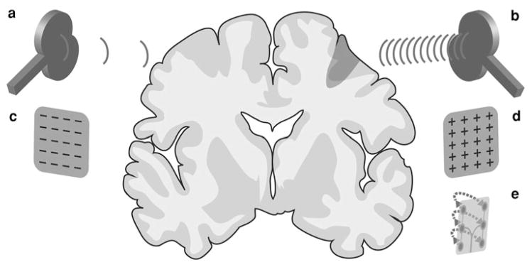
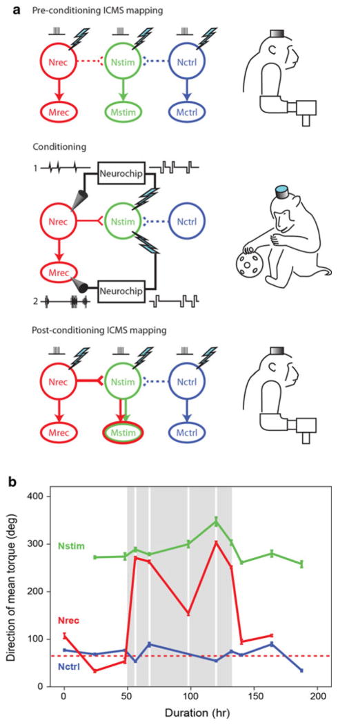
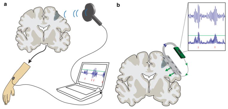
Stroke is a leading cause of disability, and the number of stroke survivors continues to rise. Traditional neurorehabilitation strategies aimed at restoring function to weakened limbs provide only modest benefit. New brain stimulation techniques designed to augment traditional neurorehabilitation hold promise for reducing the burden of stroke-related disability. Investigators discovered that repetitive transcranial magnetic stimulation (rTMS), transcranial direct current stimulation (tDCS), and epidural cortical stimulation (ECS) can enhance neural plasticity in the motor cortex post-stroke. Improved outcomes may be obtained with activity-dependent stimulation, in which brain stimulation is contingent on neural or muscular activity during normal behavior. We review the evidence for improved motor function in stroke patients treated with rTMS, tDCS, and ECS and discuss the mediating physiological mechanisms. We compare these techniques to activity-dependent stimulation, discuss the advantages of this newer strategy for stroke rehabilitation, and suggest future applications for activity-dependent brain stimulation.


Figures

Fig. 1
Fig. 1
Preprogrammed methods of brain stimulation for stroke rehabilitation. a Low-frequency (1 Hz) rTMS to contralesional M1. b High-frequency (≥3 Hz) rTMS to lesional M1. c Cathodal tDCS to contralesional M1. d Anodal tDCS to lesional M1. e Epidural cortical stimulation to lesional M1. Dark wedge-shaped region denotes lesion from cortical stroke or region impaired by subcortical stroke. Note that in practice, a and c are situated directly over contralesional M1 while b, d, and e are situated directly over lesional M1
Fig. 2
Fig. 2
Activity-dependent conditioning triggered by cortical or EMG activity in a non-human primate. a Summary of experiments demonstrating plasticity obtained with the head-fixed computer [“Neurochip”]. Preconditioning intracranial microstimulation (ICMS) trains activated distinct descending projections from each of 3 cortical sites to corresponding muscles, with monkey at rest. Conditioning during unrestrained behavior by (1) spike-triggered stimulation (Jackson et al. 2006) or (2) EMG-triggered (Lucas 2009; Lucas and Fetz 2011) stimulation induced a strengthening of horizontal connections between Nrec and Nstim. Post-conditioning ICMS now activates Mstim via strengthened horizontal projections to Nstim, as well as Mrec via the direct projection. b The direction of mean torque evoked by electrical stimulation of three M1 sites before, during, and after activity-dependent conditioning (gray regions) converted into angular degrees. M1 output effects are quantified by measuring forelimb torque responses evoked with trains of intracor-tical microstimulation (ICMS) before and after conditioning. Nrec (red)—cortical site that activates Mrec (EDC) at baseline, Nstim (green)—cortical site that activates Mstim (SUP), Nctrl (blue)—cortical site that activates Mctrl (APB). During conditioning, Mrec (EDC) muscle activity triggered intracortical stimulation at the Nstim site. Mean baseline responses evoked from Nrec stimulation illustrated with dotted red line. EDC extensor digitorum communis, SUP supinator, APB abductor pollicis brevis. Error bars SEM. Data represent the initial 50 ms of train-triggered torque responses following ICMS onset converted into angular degrees. Figures adapted from (Jackson et al. 2006) (a) and (Lucas 2009; Lucas and Fetz 2011) (b)
Fig. 3
Fig. 3
Activity-dependent methods of brain stimulation. a TMS to lesional M1 triggered by EMG activity in the paretic limb. In this schematic, the laptop computer processes EMG activity to create a triggering signal (red vertical lines) to the TMS device when the EMG signal rises above a predefined threshold (horizontal green line). b Epidural cortical stimulation (ECS) to lesional M1 triggered by neural activity in the form of high gamma band (80–120 Hz) filtered electrocorticography (ECoG) signals over hand area of M1. In this schematic, ECS is directed more laterally (green arrows) over face area of M1 in an attempt to drive vicariation. An implanted computer chip triggers ECS stimuli (red vertical lines) when rectified, high gamma band ECoG rises above a predefined threshold (horizontal green line)

References

    1. Abraham WC. Metaplasticity: tuning synapses and networks for plasticity. Nat Rev Neurosci. 2008;9:387–399. - PubMed
    1. Ackerley SJ, Stinear CM, Barber PA, Byblow WD. Combining theta burst stimulation with training after subcortical stroke. Stroke. 2010;41:1568–1572. - PubMed
    1. Adkins DL, Campos P, Quach D, et al. Epidural cortical stimulation enhances motor function after sensorimotor cortical infarcts in rats. Exp Neurol. 2006;200:356–370. - PubMed
    1. Adkins DL, Hsu JE, Jones TA. Motor cortical stimulation promotes synaptic plasticity and behavioral improvements following sensorimotor cortex lesions. Exp Neurol. 2008;212:14–28. - PMC - PubMed
    1. Adkins-Muir DL, Jones TA. Cortical electrical stimulation combined with rehabilitative training: enhanced functional recovery and dendritic plasticity following focal cortical ischemia in rats. Neurol Res. 2003;25:780–788. - PubMed

Publication types

MeSH terms