. 2013 Mar;132(1):96-106.
doi: 10.1093/toxsci/kfs328. Epub 2012 Nov 28.

Exposure to triclosan augments the allergic response to ovalbumin in a mouse model of asthma

Affiliations

Exposure to triclosan augments the allergic response to ovalbumin in a mouse model of asthma

Stacey E Anderson et al. Toxicol Sci. 2013 Mar.

Abstract

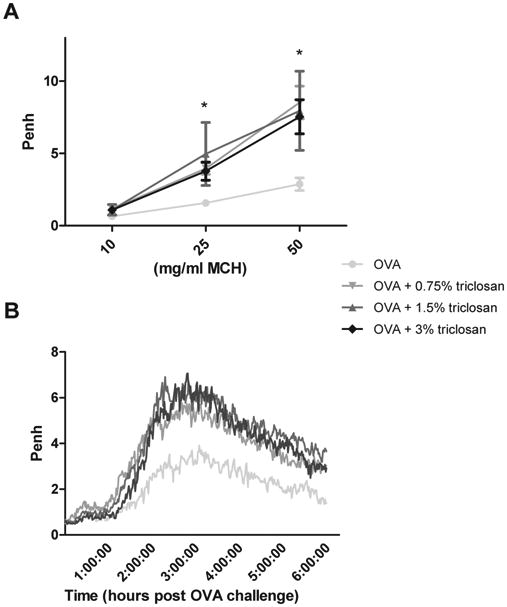
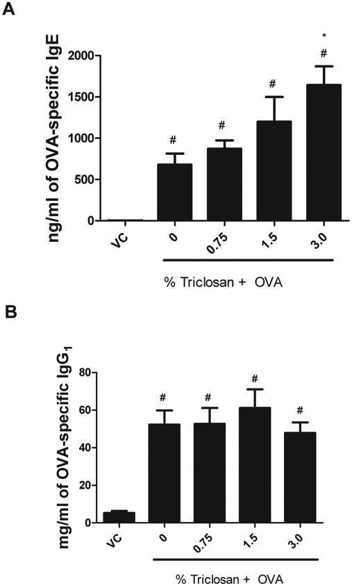
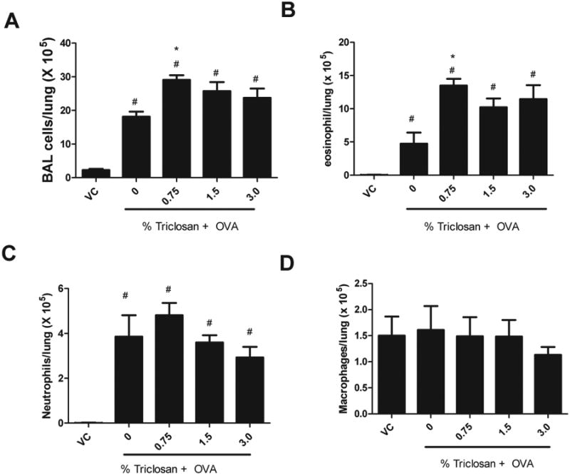
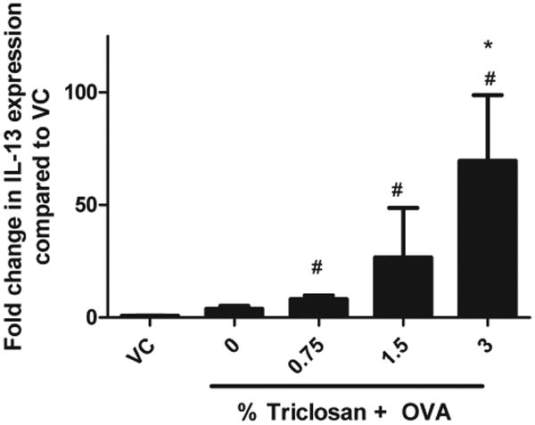
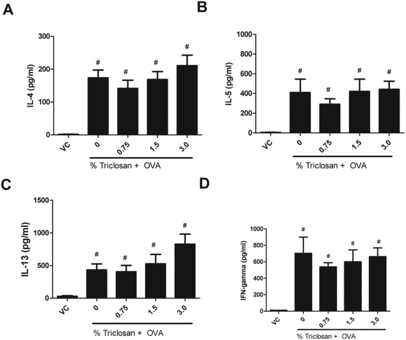
During the last decade, there has been a remarkable and unexplained increase in the prevalence of asthma. These studies were conducted to investigate the role of dermal exposure to triclosan, an endocrine-disrupting compound, on the hypersensitivity response to ovalbumin (OVA) in a murine model of asthma. Triclosan has had widespread use in the general population as an antibacterial and antifungal agent and is commonly found in consumer products such as soaps, deodorants, toothpastes, shaving creams, mouthwashes, and cleaning supplies. For these studies, BALB/c mice were exposed dermally to concentrations of triclosan ranging from 0.75 to 3% (0.375-1.5mg/mouse/day) for 28 consecutive days. Concordantly, mice were ip injected with OVA (0.9 µg) and aluminum hydroxide (0.5mg) on days 1 and 10 and challenged with OVA (125 µg) by pharyngeal aspiration on days 19 and 27. Compared with the animals exposed to OVA alone, increased spleen weights, OVA-specific IgE, interleukin-13 cytokine levels, and numbers of lung eosinophils were demonstrated when mice were coexposed to OVA and triclosan. Statistically significant increases in OVA-specific and nonspecific airway hyperreactivity were observed for all triclosan coexposed groups compared with the vehicle and OVA controls. In these studies, exposure to triclosan alone was not demonstrated to be allergenic; however, coexposure with a known allergen resulted in enhancement of the hypersensitivity response to that allergen, suggesting that triclosan exposure may augment the allergic responses to other environmental allergens.


Figures

Fig. 1
Fig. 1
Effect of coexposure to triclosan and OVA on airway hyperreactivity. Nonspecific (A) and specific (B) airway hyperreactivity were evaluated in mice (eight per group) that were dermally exposed to triclosan for 28 days and sensitized with OVA on days 1 and 7 of the experiment and then challenged with OVA via pharyngeal aspiration on days 19 and 27. Nonspecific AHR was evaluated on day 21, and specific AHR was evaluated on day 27 immediately following pulmonary challenge. Each point/line represents mean values (baseline subtracted) ± SE of eight mice per group. Levels of statistical significance are designated as * (p < 0.05) for all coexposure groups compared with the OVA control. The data graphed are the average Penh value plotted over the 6-h collection period for the OVA-specific airway response.
Fig. 2
Fig. 2
OVA-specific antibody production following coexposure to triclosan and OVA. Analysis of OVA-specific (A) IgE and (B) IgG1, following exposure to triclosan and OVA, was measured using an ELISA. Bars represent mean ± SE for each group of eight mice. Statistical significance (p ≤ 0.05) is designated as * compared with the OVA control or # compared with the VC.
Fig. 3
Fig. 3
Effect of coexposure to triclosan and OVA on BAL infiltrates. Absolute numbers of (A) total BAL cells, (B) eosinophils, (C) neutrophils, and (D) macrophages evaluated in mice following exposure to triclosan + OVA. Error bars represent mean ± SE of seven mice per group. Statistical significance (p ≤ 0.05) is designated as * compared with the OVA control or # compared with the VC.
Fig. 4
Fig. 4
Lung histopathology following coexposure to triclosan and OVA. Photomicrographs represent (A) lung of a VC mouse (bar = 50 μm); (B) lung of a mouse exposed to OVA and 3% triclosan demonstrating peribronchiolar, vascular, and perivascular infiltrates with extension into alveolar spaces (bar = 50 μm); (C) higher magnifications of the lung from the mouse in B showing mucous metaplasia and peribronchiolar plasma cells and eosinophils (bar = 20 μm); (D) lung of a mouse exposed to OVA alone showing perivascular and peribronchiolar inflitrates with extension into alveolar spaces (bar = 50 μm).
Fig.5
Fig.5
Effect of coexposure to triclosan and OVA on IL-13 mRNA cytokine production by the Lung. IL-13 mRNA expression was evaluated in mice following coexposure to triclosan + OVA. Error bars represent mean ± SE of four mice per group. Statistical significance (p ≤ 0.05) is designated as * compared with the OVA control or # compared with the VC.
Fig. 6
Fig. 6
Effect of coexposure to triclosan and OVA on cytokine production by the tracheobronchial lymph node. (A) IL-4, (B) IL-5, (C) IL-13, and (D) IFN-γ protein expression were evaluated by ELISA in ex vivo stimulated lymph node cells following coexposure to triclosan + OVA. Error bars represent mean ± SE of eight mice per group. Statistical significance (p ≤ 0.05) is designated as # compared with the VC.

Similar articles

Cited by

References

    1. Adolfsson-Erici M, Pettersson M, Parkkonen J, Sturve J. Triclosan, a commonly used bactericide found in human milk and in the aquatic environment in Sweden. Chemosphere. 2002;46:1485–1489. - PubMed
    1. Ahn KC, Zhao B, Chen J, Cherednichenko G, Sanmarti E, Denison MS, Lasley B, Pessah IN, Kültz D, Chang DP, et al. In vitro biologic activities of the antimicrobials triclocarban, its analogs, and triclosan in bioassay screens: Receptor-based bioassay screens. Environ Health Perspect. 2008;116:1203–1210. - PMC - PubMed
    1. Barros SP, Wirojchanasak S, Barrow DA, Panagakos FS, Devizio W, Offenbacher S. Triclosan inhibition of acute and chronic inflammatory gene pathways. J Clin Periodontol. 2010;37:412–418. - PubMed
    1. Black JG, Howes D, Rutherford T. Percutaneous absorption and metabolism of Irgasan DP300. Toxicology. 1975;3:33–47. - PubMed
    1. Bracci L, Vukcevic M, Spagnoli G, Ducreux S, Zorzato F, Treves S. Ca2+ signaling through ryanodine receptor 1 enhances maturation and activation of human dendritic cells. J Cell Sci. 2007;120(Pt 13):2232–2240. - PubMed

MeSH terms