

Osteoconductive phosphoserine-modified poly({varepsilon}-lysine) dendrons: synthesis, titanium oxide surface functionalization and response of osteoblast-like cell lines
- PMID: 23193106
- PMCID: PMC3565693
- DOI: 10.1098/rsif.2012.0765
Osteoconductive phosphoserine-modified poly({varepsilon}-lysine) dendrons: synthesis, titanium oxide surface functionalization and response of osteoblast-like cell lines
Abstract
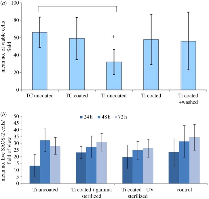
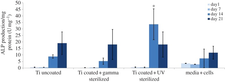
The lack of direct bonding between the surface of an implant and the mineralized bony tissue is among the main causes of aseptic loosening in titanium-based implants. Surface etching and ceramic coatings have led to improved osteointegration, but their clinical performance is still limited either by partial bonding or by coating delamination. In this work, a solid-phase synthesis method has been optimized to produce poly(ε-lysine) dendrons, the outermost branching generation of which is functionalized by phosphoserine (PS), a known catalyst of the biomineralization process. The dendrons were deposited onto etched titanium oxide surfaces as a near-to-monolayer film able to induce the formation of a homogeneous calcium phosphate phase in a simulated body fluid over 3 days. The dendron films also stimulated MG63 and SAOS-2 osteoblast-like cells to proliferate at a rate significantly higher than etched titanium, with SAOS-2 also showing a higher degree of differentiation over 14 days. PS-tethered dendron films were not affected by various sterilization methods and UV treatment appeared to improve the cell substrate potential of these films, thus suggesting their potential as a surface functionalization method for bone implants.
Figures
Similar articles
-
Osseointegration of titanium implants functionalised with phosphoserine-tethered poly(epsilon-lysine) dendrons: a comparative study with traditional surface treatments in sheep.J Mater Sci Mater Med. 2015 Feb;26(2):87. doi: 10.1007/s10856-015-5433-3. Epub 2015 Feb 3. J Mater Sci Mater Med. 2015. PMID: 25644101
-
Biomimetic coating with phosphoserine-tethered poly(epsilon-lysine) dendrons on titanium surfaces enhances Wnt and osteoblastic differentiation.Clin Oral Implants Res. 2014 Feb;25(2):e133-9. doi: 10.1111/clr.12075. Epub 2012 Dec 5. Clin Oral Implants Res. 2014. PMID: 23210635
-
Complexation of Injectable Biphasic Calcium Phosphate with Phosphoserine-Presenting Dendrons with Enhanced Osteoregenerative Properties.ACS Appl Mater Interfaces. 2020 Aug 26;12(34):37873-37884. doi: 10.1021/acsami.0c09004. Epub 2020 Aug 13. ACS Appl Mater Interfaces. 2020. PMID: 32687309
-
Supramolecular structures from dendrons and dendrimers.Adv Drug Deliv Rev. 2005 Dec 14;57(15):2238-70. doi: 10.1016/j.addr.2005.09.015. Epub 2005 Nov 28. Adv Drug Deliv Rev. 2005. PMID: 16310885 Review.
-
Genetic potential of interfacial guided osteogenesis in implant devices.Dent Mater J. 2000 Jun;19(2):99-132. doi: 10.4012/dmj.19.99. Dent Mater J. 2000. PMID: 11219100 Review.
Cited by
-
Poly(ε-lysine) dendrons as modulators of quorum sensing in Pseudomonas aeruginosa.J Mater Sci Mater Med. 2015 May;26(5):176. doi: 10.1007/s10856-015-5508-1. Epub 2015 Apr 17. J Mater Sci Mater Med. 2015. PMID: 25893386
-
Dendritic Polymers in Tissue Engineering: Contributions of PAMAM, PPI PEG and PEI to Injury Restoration and Bioactive Scaffold Evolution.Pharmaceutics. 2023 Feb 4;15(2):524. doi: 10.3390/pharmaceutics15020524. Pharmaceutics. 2023. PMID: 36839847 Free PMC article. Review.
-
A Novel Class of Injectable Bioceramics that Glue Tissues and Biomaterials.Materials (Basel). 2018 Dec 7;11(12):2492. doi: 10.3390/ma11122492. Materials (Basel). 2018. PMID: 30544596 Free PMC article.
-
Adhesive Cements That Bond Soft Tissue Ex Vivo.Materials (Basel). 2019 Aug 3;12(15):2473. doi: 10.3390/ma12152473. Materials (Basel). 2019. PMID: 31382566 Free PMC article.
-
Osseointegration of titanium implants functionalised with phosphoserine-tethered poly(epsilon-lysine) dendrons: a comparative study with traditional surface treatments in sheep.J Mater Sci Mater Med. 2015 Feb;26(2):87. doi: 10.1007/s10856-015-5433-3. Epub 2015 Feb 3. J Mater Sci Mater Med. 2015. PMID: 25644101
References
-
- Mendonça G, Mendonça DBS, Aragão FJL, Cooper LF. 2008. Advancing dental implant surface technology: from micron to nanotopography. Biomaterials 29, 3822–383510.1016/j.biomaterials.2008.05.012 (doi:10.1016/j.biomaterials.2008.05.012) - DOI - DOI - PubMed
-
- Cao W, Hench LL. 1996. Bioactive materials. Ceram. Int. 22, 493–50710.1016/0272-8842(95)00126-3 (doi:10.1016/0272-8842(95)00126-3) - DOI - DOI
-
- Orsini G, Assenza B, Scarano A, Piattelli M, Piattelli A. 2000. Surface analysis of machined versus sandblasted and acid-etched titanium implants. Int. J. Oral Maxillofac. Implants 15, 779–84 - PubMed
-
- Habibovic P, Barrere F, van Blitterswijk CA, DeGroot K, Layrolle P. 2002. Biomimetic hydroxyapatite coating on metal implants. J. Am. Ceram. Soc. 85, 517–52210.1111/j.1151-2916.2002.tb00126.x (doi:10.1111/j.1151-2916.2002.tb00126.x) - DOI - DOI
-
- Wen J, Leng Y, Chen J, Zhang C. 2000. Chemical gradient in plasma-sprayed HA coatings. Biomaterials 21, 1339–134310.1016/S0142-9612(99)00273-2 (doi:10.1016/S0142-9612(99)00273-2) - DOI - DOI - PubMed
Publication types
MeSH terms
Substances
LinkOut - more resources
Full Text Sources
Other Literature Sources
