. 2013 Mar;62(3):896-904.
doi: 10.2337/db12-0929. Epub 2012 Nov 28.

PTPN22 silencing in the NOD model indicates the type 1 diabetes-associated allele is not a loss-of-function variant

Affiliations

PTPN22 silencing in the NOD model indicates the type 1 diabetes-associated allele is not a loss-of-function variant

Peilin Zheng et al. Diabetes. 2013 Mar.

Abstract

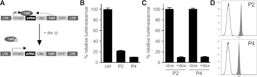
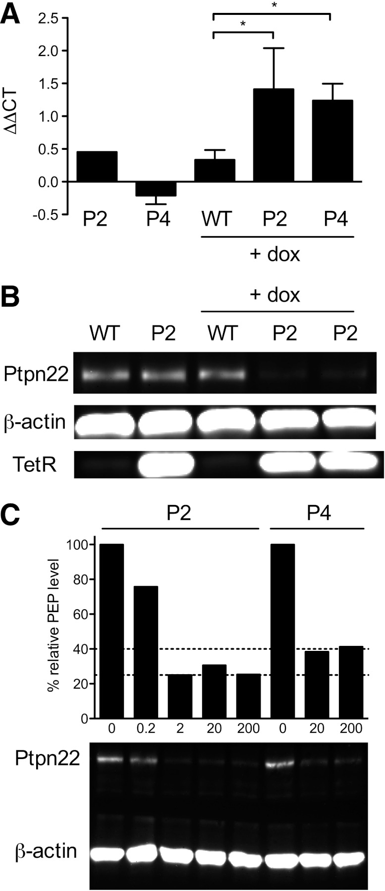
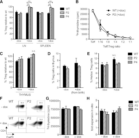
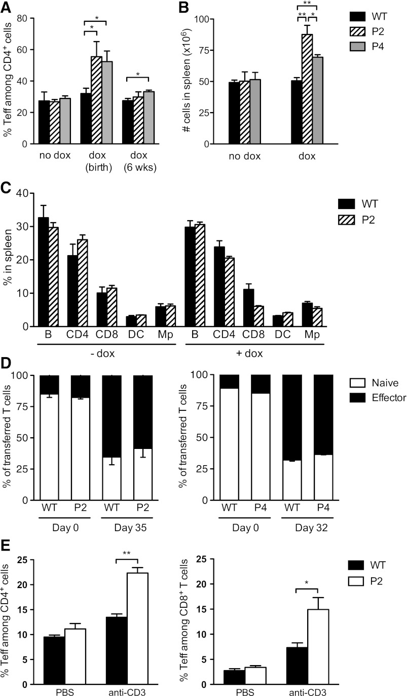
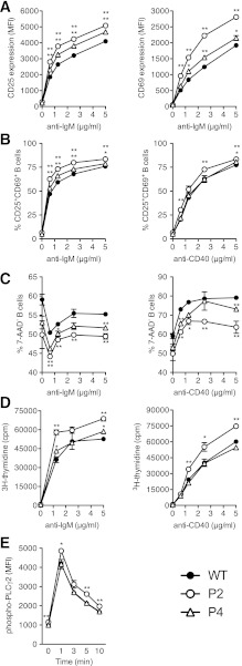
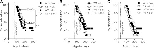
PTPN22 encodes the lymphoid tyrosine phosphatase (LYP) and is the second strongest non-HLA genetic risk factor for type 1 diabetes. The PTPN22 susceptibility allele generates an LYP variant with an arginine-to-tryptophan substitution at position 620 (R620W) that has been reported by several studies to impart a gain of function. However, a recent report investigating both human cells and a knockin mouse model containing the R620W homolog suggested that this variation causes faster protein degradation. Whether LYP R620W is a gain- or loss-of-function variant, therefore, remains controversial. To address this issue, we generated transgenic NOD mice (nonobese diabetic) in which Ptpn22 can be inducibly silenced by RNA interference. We found that Ptpn22 silencing in the NOD model replicated many of the phenotypes observed in C57BL/6 Ptpn22 knockout mice, including an increase in regulatory T cells. Notably, loss of Ptpn22 led to phenotypic changes in B cells opposite to those reported for the human susceptibility allele. Furthermore, Ptpn22 knockdown did not increase the risk of autoimmune diabetes but, rather, conferred protection from disease. Overall, to our knowledge, this is the first functional study of Ptpn22 within a model of type 1 diabetes, and the data do not support a loss of function for the PTPN22 disease variant.


Figures

FIG. 1.
FIG. 1.
Generation of inducible Ptpn22 KD mice. A: Schematic representation of the inducible construct used for transgenesis. Long terminal repeats (LTRs) flank a cassette containing a tetracycline-regulated H1 promoter (H1-tetO) driving shRNA expression and a ubiquitin (Ubq) promoter driving expression of the TetR and GFP. Addition of doxycycline (dox) derepresses the H1 promoter and induces shRNA expression. B: The silencing efficiency of shRNAs P2 and P4 was validated by cotransfection into 293-F cells of a Ptpn22 luciferase reporter and lentiviral vectors containing constitutively expressed P2 or P4 shRNA. C: 293-F cells were infected with inducible P2 or P4 lentivirus, cultured in the presence or absence of dox, and transfected with the Ptpn22 luciferase reporter to test for inducibility of the KD constructs. Data in B and C are representative of two independent experiments. D: Representative GFP expression in blood lymphocytes from transgenic P2 and P4 mice. Forward- and side-scatter characteristics were used to gate on the lymphocytic population. Open histogram, nontransgenic; solid histogram, transgenic.
FIG. 2.
FIG. 2.
Validation of Ptpn22 silencing in transgenic P2 and P4 mice. A: Quantitation of Ptpn22 mRNA levels in lymphocytes from untreated P2 and P4 mice and from doxycycline (dox)-treated WT, P2, and P4 mice relative to Ptpn22 expression in untreated WT mice. ΔΔCTs (ΔCT of sample – ΔCT untreated WT such that an increase in ΔΔCT indicates lower mRNA levels) are shown. Data were averaged from four independent experiments. B: Quantitation of PEP (Ptpn22) protein by Western blotting in lymphocytes from untreated or dox-treated (200 μg/mL) WT and P2 mice. TetR was also assayed to verify transgene expression. C: Quantitation of PEP protein in lymphocytes after exposure to increasing doses of dox (μg/mL) for both P2 and P4 mice. Data in B and C are representative of six and two similar experiments, respectively. All mice were 8–12 weeks old and had received dox for 10–14 days. *P < 0.05. CT, cycle threshold.
FIG. 3.
FIG. 3.
Ptpn22 silencing increases peripheral but not thymic Treg-cell numbers. A: The percentage of FoxP3+CD4+ T cells in lymph nodes (LNs) or spleen of untreated or doxycycline (dox)-treated (for 4 weeks from the age of 8 weeks) WT, P2, and P4 mice relative to untreated WT (LN 11.23%, spleen 16.7%). Data were averaged from three mice per group and are representative of 13 similar experiments. B: Proliferation of WT CD4+CD25 T cells in response to anti-CD3 stimulation was measured in the presence or absence of CD4+CD25+ T cells from WT or P2 mice treated with dox for 2 weeks from the age of 8 weeks. Data are mean ± SEM of triplicates and are representative of three independent experiments. C: The percentage FoxP3+CD4+CD8 T cells in the thymus of untreated or dox-treated (as in A) WT, P2, and P4 mice relative to untreated WT (7.27%). D: The percentage FoxP3+CD4+CD8 T cells in the thymus of untreated or dox-treated (for 2 months from birth) WT, P2, and P4 mice. E: The percentage of Helios-negative cells among FoxP3+CD4+ T cells in lymph nodes from untreated or dox-treated (as in A) WT, P2, and P4 mice. Data in C, D, and E were averaged from three to four mice per group and are representative of five (C and E) and two (D) similar experiments. F: CD62LhiCD4+ T cells from untreated or dox-treated (for 2 weeks from 8 weeks of age) WT and P2 mice were activated in the presence of transforming growth factor-β (with or without dox) for 3 days before measuring FoxP3 expression. Data are representative of two independent experiments. G and H: Proliferation at 72 h (G) and fold numerical expansion after 10 days in culture (H) of Treg cells from dox-treated (for 2 weeks from 6 weeks of age) or untreated WT, P2, and P4 mice after stimulation with CD3- and CD28-antibody–coated beads; data are mean ± SEM of triplicate measurements. **P < 0.01. NS, not significant at P > 0.05.
FIG. 4.
FIG. 4.
Time of treatment determines the effect of Ptpn22 silencing on Teff-cell differentiation. A: The percentage CD44hiCD62Llo (Teff) cells within the CD4+ T-cell compartment were quantified in WT, P2, and P4 mice that had either been untreated, treated for 2 months from birth, or treated for 3 months from the age of 6 weeks. Data for CD8+ T cells were similar (not shown). Data are averaged (mean ± SEM) for two to six mice per group and are representative of two (doxycycline [dox] from birth) and six (dox from 6 weeks) similar experiments. B: Cellularity of spleens from WT, P2, and P4 mice untreated or treated with dox for 4–5 months from the age of 10 weeks averaged (mean ± SEM) for 14 WT, 5 P2, and 8 P4 mice (untreated groups) and 19 WT, 10 P2, and 9 P4 mice (treated groups). C: Relative distribution of B cells, CD4 and CD8 T cells, dendritic cells (DC), and macrophages (Mp) in the spleen of WT and P2 mice either untreated or treated from the age of 4 weeks for a duration of 4 months. Data are averaged (mean ± SEM) from five mice per group and representative of four similar experiments. D: Quantification of naïve (CD44loCD62Lhi) and effector (CD44hiCD62Llo) cells in mixed WT and P2 or WT and P4 CD4+ T-cell populations before and after (32–35 days) transfer into NOD.scid mice. WT and transgenic cells were identified on the basis of GFP expression. E: The percentage of CD44hiCD62Llo (Teff) cells was quantified in CD4+ and CD8+ T cells from WT and P2 mice (pretreated with dox for 10 days) 2 days after injection with PBS only or anti-CD3 antibody 10 μg. Data in D and E are averaged from three to four mice per group and representative of two independent experiments each. *P < 0.05; **P < 0.01.
FIG. 5.
FIG. 5.
Ptpn22 silencing increases activation and decreases survival of transgenic B cells. B cells from WT, P2, and P4 mice treated with doxycycline for 4 weeks from 8 weeks of age were stimulated with anti-IgM (AE) or anti-CD40 (BD) at the indicated concentration. Mean fluorescence intensity (MFI) of CD25 and CD69 staining (A), percentage of CD25+CD69+ cells (B), and percentage of 7-aminoactinomycin D negative (7-AAD) (nonapoptotic) cells (C) were quantified 24 h after stimulation. Proliferation was measured by 3H-thymidine incorporation 48 h poststimulation (D). The phosphorylation of PLCγ2 was measured at a single concentration of anti-IgM (40 μg/mL) at the indicated time poststimulation (E). Data are representative of four (AC), seven (D), and two (E) independent experiments. Data are mean ± SEM of triplicate measurements. *P < 0.05; **P < 0.01.
FIG. 6.
FIG. 6.
P2 but not P4 mice are protected from autoimmune diabetes by Ptpn22 silencing. Female P2 (A) and P4 (B and C) transgenic mice and their WT littermates were either left untreated or treated with doxycycline (dox) (200 μg/mL drinking water) from the age of 10 weeks (A and B) or 4 weeks (C). The dashed line indicates start of treatment. A: WT – dox (n = 20), P2 – dox (n = 31), WT + dox (n = 15), P2 + dox (n = 18). B: WT – dox (n = 36), P4 – dox (n = 29), WT + dox (n = 20), P4 + dox n = 15. C: WT – dox n = 36, P4 – dox n = 29, WT + dox n = 32, P4 + dox n = 34. Log-rank test for A: WT – dox versus P2 – dox P = 0.26, WT + dox versus P2 + dox P = 0.006, P2 – dox versus P2 + dox P = 0.0418 (*). NS, not significant.

Similar articles

Cited by

References

    1. Cohen S, Dadi H, Shaoul E, Sharfe N, Roifman CM. Cloning and characterization of a lymphoid-specific, inducible human protein tyrosine phosphatase, Lyp. Blood 1999;93:2013–2024 - PubMed
    1. Cloutier JF, Veillette A. Cooperative inhibition of T-cell antigen receptor signaling by a complex between a kinase and a phosphatase. J Exp Med 1999;189:111–121 - PMC - PubMed
    1. Rieck M, Arechiga A, Onengut-Gumuscu S, Greenbaum C, Concannon P, Buckner JH. Genetic variation in PTPN22 corresponds to altered function of T and B lymphocytes. J Immunol 2007;179:4704–4710 - PubMed
    1. Bottini N, Musumeci L, Alonso A, et al. A functional variant of lymphoid tyrosine phosphatase is associated with type I diabetes. Nat Genet 2004;36:337–338 - PubMed
    1. Kyogoku C, Langefeld CD, Ortmann WA, et al. Genetic association of the R620W polymorphism of protein tyrosine phosphatase PTPN22 with human SLE. Am J Hum Genet 2004;75:504–507 - PMC - PubMed

Publication types

MeSH terms

Substances