

Roles of yeast eIF2α and eIF2β subunits in the binding of the initiator methionyl-tRNA
- PMID: 23193270
- PMCID: PMC3553985
- DOI: 10.1093/nar/gks1180
Roles of yeast eIF2α and eIF2β subunits in the binding of the initiator methionyl-tRNA
Abstract
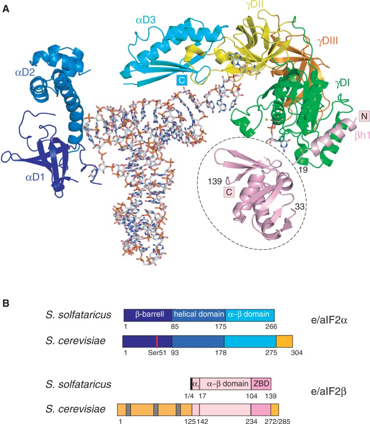
Heterotrimeric eukaryotic/archaeal translation initiation factor 2 (e/aIF2) binds initiator methionyl-tRNA and plays a key role in the selection of the start codon on messenger RNA. tRNA binding was extensively studied in the archaeal system. The γ subunit is able to bind tRNA, but the α subunit is required to reach high affinity whereas the β subunit has only a minor role. In Saccharomyces cerevisiae however, the available data suggest an opposite scenario with β having the most important contribution to tRNA-binding affinity. In order to overcome difficulties with purification of the yeast eIF2γ subunit, we designed chimeric eIF2 by assembling yeast α and β subunits to archaeal γ subunit. We show that the β subunit of yeast has indeed an important role, with the eukaryote-specific N- and C-terminal domains being necessary to obtain full tRNA-binding affinity. The α subunit apparently has a modest contribution. However, the positive effect of α on tRNA binding can be progressively increased upon shortening the acidic C-terminal extension. These results, together with small angle X-ray scattering experiments, support the idea that in yeast eIF2, the tRNA molecule is bound by the α subunit in a manner similar to that observed in the archaeal aIF2-GDPNP-tRNA complex.
Figures
Similar articles
-
tRNA binding properties of eukaryotic translation initiation factor 2 from Encephalitozoon cuniculi.Biochemistry. 2010 Oct 12;49(40):8680-8. doi: 10.1021/bi1009166. Epub 2010 Sep 15. Biochemistry. 2010. PMID: 20822097
-
Initiation factor eIF2γ promotes eIF2-GTP-Met-tRNAi(Met) ternary complex binding to the 40S ribosome.Nat Struct Mol Biol. 2011 Oct 16;18(11):1227-34. doi: 10.1038/nsmb.2133. Nat Struct Mol Biol. 2011. PMID: 22002225 Free PMC article.
-
Structure of the ternary initiation complex aIF2-GDPNP-methionylated initiator tRNA.Nat Struct Mol Biol. 2012 Mar 25;19(4):450-4. doi: 10.1038/nsmb.2259. Nat Struct Mol Biol. 2012. PMID: 22447243
-
Eukaryotic and archaeal translation initiation factor 2: a heterotrimeric tRNA carrier.FEBS Lett. 2010 Jan 21;584(2):405-12. doi: 10.1016/j.febslet.2009.11.002. FEBS Lett. 2010. PMID: 19896944 Review.
-
Eukaryotic initiator tRNA: finely tuned and ready for action.FEBS Lett. 2010 Jan 21;584(2):396-404. doi: 10.1016/j.febslet.2009.11.047. FEBS Lett. 2010. PMID: 19925799 Free PMC article. Review.
Cited by
-
eIF2 interactions with initiator tRNA and eIF2B are regulated by post-translational modifications and conformational dynamics.Cell Discov. 2015 Aug 11;1:15020. doi: 10.1038/celldisc.2015.20. eCollection 2015. Cell Discov. 2015. PMID: 27462419 Free PMC article.
-
Role of aIF5B in archaeal translation initiation.Nucleic Acids Res. 2022 Jun 24;50(11):6532-6548. doi: 10.1093/nar/gkac490. Nucleic Acids Res. 2022. PMID: 35694843 Free PMC article.
-
eIF2B is a decameric guanine nucleotide exchange factor with a γ2ε2 tetrameric core.Nat Commun. 2014 May 23;5:3902. doi: 10.1038/ncomms4902. Nat Commun. 2014. PMID: 24852487 Free PMC article.
-
Cryo-EM study of start codon selection during archaeal translation initiation.Nat Commun. 2016 Nov 7;7:13366. doi: 10.1038/ncomms13366. Nat Commun. 2016. PMID: 27819266 Free PMC article.
-
eIF2A, an initiator tRNA carrier refractory to eIF2α kinases, functions synergistically with eIF5B.Cell Mol Life Sci. 2018 Dec;75(23):4287-4300. doi: 10.1007/s00018-018-2870-4. Epub 2018 Jul 17. Cell Mol Life Sci. 2018. PMID: 30019215 Free PMC article.
References
Publication types
MeSH terms
Substances
LinkOut - more resources
Full Text Sources
Molecular Biology Databases
