

Computerized detection of lung nodules by means of "virtual dual-energy" radiography
- PMID: 23193306
- PMCID: PMC4283823
- DOI: 10.1109/TBME.2012.2226583
Computerized detection of lung nodules by means of "virtual dual-energy" radiography
Abstract
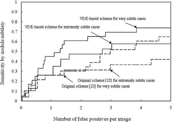
Major challenges in current computer-aided detection (CADe) schemes for nodule detection in chest radiographs (CXRs) are to detect nodules that overlap with ribs and/or clavicles and to reduce the frequent false positives (FPs) caused by ribs. Detection of such nodules by a CADe scheme is very important, because radiologists are likely to miss such subtle nodules. Our purpose in this study was to develop a CADe scheme with improved sensitivity and specificity by use of "virtual dual-energy" (VDE) CXRs where ribs and clavicles are suppressed with massive-training artificial neural networks (MTANNs). To reduce rib-induced FPs and detect nodules overlapping with ribs, we incorporated the VDE technology in our CADe scheme. The VDE technology suppressed rib and clavicle opacities in CXRs while maintaining soft-tissue opacity by use of the MTANN technique that had been trained with real dual-energy imaging. Our scheme detected nodule candidates on VDE images by use of a morphologic filtering technique. Sixty morphologic and gray-level-based features were extracted from each candidate from both original and VDE CXRs. A nonlinear support vector classifier was employed for classification of the nodule candidates. A publicly available database containing 140 nodules in 140 CXRs and 93 normal CXRs was used for testing our CADe scheme. All nodules were confirmed by computed tomography examinations, and the average size of the nodules was 17.8 mm. Thirty percent (42/140) of the nodules were rated "extremely subtle" or "very subtle" by a radiologist. The original scheme without VDE technology achieved a sensitivity of 78.6% (110/140) with 5 (1165/233) FPs per image. By use of the VDE technology, more nodules overlapping with ribs or clavicles were detected and the sensitivity was improved substantially to 85.0% (119/140) at the same FP rate in a leave-one-out cross-validation test, whereas the FP rate was reduced to 2.5 (583/233) per image at the same sensitivity level as the original CADe scheme obtained (Difference between the specificities of the original and the VDE-based CADe schemes was statistically significant). In particular, the sensitivity of our VDE-based CADe scheme for subtle nodules (66.7% = 28/42) was statistically significantly higher than that of the original CADe scheme (57.1% = 24/42). Therefore, by use of VDE technology, the sensitivity and specificity of our CADe scheme for detection of nodules, especially subtle nodules, in CXRs were improved substantially.
Figures


Similar articles
-
Development and evaluation of a computer-aided diagnostic scheme for lung nodule detection in chest radiographs by means of two-stage nodule enhancement with support vector classification.Med Phys. 2011 Apr;38(4):1844-58. doi: 10.1118/1.3561504. Med Phys. 2011. PMID: 21626918 Free PMC article.
-
Image-processing technique for suppressing ribs in chest radiographs by means of massive training artificial neural network (MTANN).IEEE Trans Med Imaging. 2006 Apr;25(4):406-16. doi: 10.1109/TMI.2006.871549. IEEE Trans Med Imaging. 2006. PMID: 16608057
-
Separation of bones from chest radiographs by means of anatomically specific multiple massive-training ANNs combined with total variation minimization smoothing.IEEE Trans Med Imaging. 2014 Feb;33(2):246-57. doi: 10.1109/TMI.2013.2284016. Epub 2013 Oct 11. IEEE Trans Med Imaging. 2014. PMID: 24132005
-
Is there an advantage to using computer aided detection for the early detection of pulmonary nodules within chest X-Ray imaging?Radiography (Lond). 2020 Aug;26(3):e170-e178. doi: 10.1016/j.radi.2020.01.002. Epub 2020 Jan 29. Radiography (Lond). 2020. PMID: 32052750 Review.
-
Research progress of computer aided diagnosis system for pulmonary nodules in CT images.J Xray Sci Technol. 2020;28(1):1-16. doi: 10.3233/XST-190581. J Xray Sci Technol. 2020. PMID: 31815727 Review.
Cited by
-
Segmentation and classification on chest radiography: a systematic survey.Vis Comput. 2023;39(3):875-913. doi: 10.1007/s00371-021-02352-7. Epub 2022 Jan 8. Vis Comput. 2023. PMID: 35035008 Free PMC article.
-
Soft Tissue/Bone Decomposition of Conventional Chest Radiographs Using Nonparametric Image Priors.Appl Bionics Biomech. 2019 Jun 24;2019:9806464. doi: 10.1155/2019/9806464. eCollection 2019. Appl Bionics Biomech. 2019. PMID: 31341514 Free PMC article.
-
Overview of deep learning in medical imaging.Radiol Phys Technol. 2017 Sep;10(3):257-273. doi: 10.1007/s12194-017-0406-5. Epub 2017 Jul 8. Radiol Phys Technol. 2017. PMID: 28689314 Review.
-
A parameterized logarithmic image processing method with Laplacian of Gaussian filtering for lung nodule enhancement in chest radiographs.Med Biol Eng Comput. 2016 Nov;54(11):1793-1806. doi: 10.1007/s11517-016-1469-x. Epub 2016 Mar 25. Med Biol Eng Comput. 2016. PMID: 27016368
-
Separation of bones from soft tissue in chest radiographs: Anatomy-specific orientation-frequency-specific deep neural network convolution.Med Phys. 2019 May;46(5):2232-2242. doi: 10.1002/mp.13468. Epub 2019 Mar 28. Med Phys. 2019. PMID: 30848498 Free PMC article.
References
-
- American Cancer Society . American Cancer Society Complete Guide to Complementary & Alternative Cancer Therapies. 2nd ed American Cancer Society; Atlanta, GA: 2009.
-
- Henschke CI, McCauley DI, Yankelevitz DF, Naidich DP, McGuinness G, Miettinen OS, Libby DM, Pasmantier MW, Koizumi J, Altorki NK, Smith JP. Early lung cancer action project: Overall design and findings from baseline screening. Lancet. 1999 Jul 10;354:99–105. - PubMed
-
- Murphy GP, Lawrence W, Lenhard RE, American Cancer Society . American Cancer Society Textbook of Clinical Oncology. 2nd ed. American Cancer Society; Atlanta, GA.: 1995.
-
- Zhao H, Lo SC, Freedman M, Wang Y. presented at the Proc. SPIE Med. Imag.: Image Process. San Diego, CA: 2002. Enhanced lung cancer detection in temporal subtraction chest radiography using directional edge filtering techniques.
-
- Austin JH, Romney BM, Goldsmith LS. Missed bronchogenic carcinoma: Radiographic findings in 27 patients with a potentially resectable lesion evident in retrospect. Radiology. 1992 Jan;182:115–122. - PubMed
Publication types
MeSH terms
Grants and funding
LinkOut - more resources
Full Text Sources
Other Literature Sources
Medical
Miscellaneous
