. 2013 Jan;53(1):97-110.
doi: 10.1016/j.jbior.2012.09.005. Epub 2012 Sep 13.

Dynamic steps in receptor tyrosine kinase mediated activation of class IA phosphoinositide 3-kinases (PI3K) captured by H/D exchange (HDX-MS)

Affiliations

Dynamic steps in receptor tyrosine kinase mediated activation of class IA phosphoinositide 3-kinases (PI3K) captured by H/D exchange (HDX-MS)

John E Burke et al. Adv Biol Regul. 2013 Jan.

Abstract

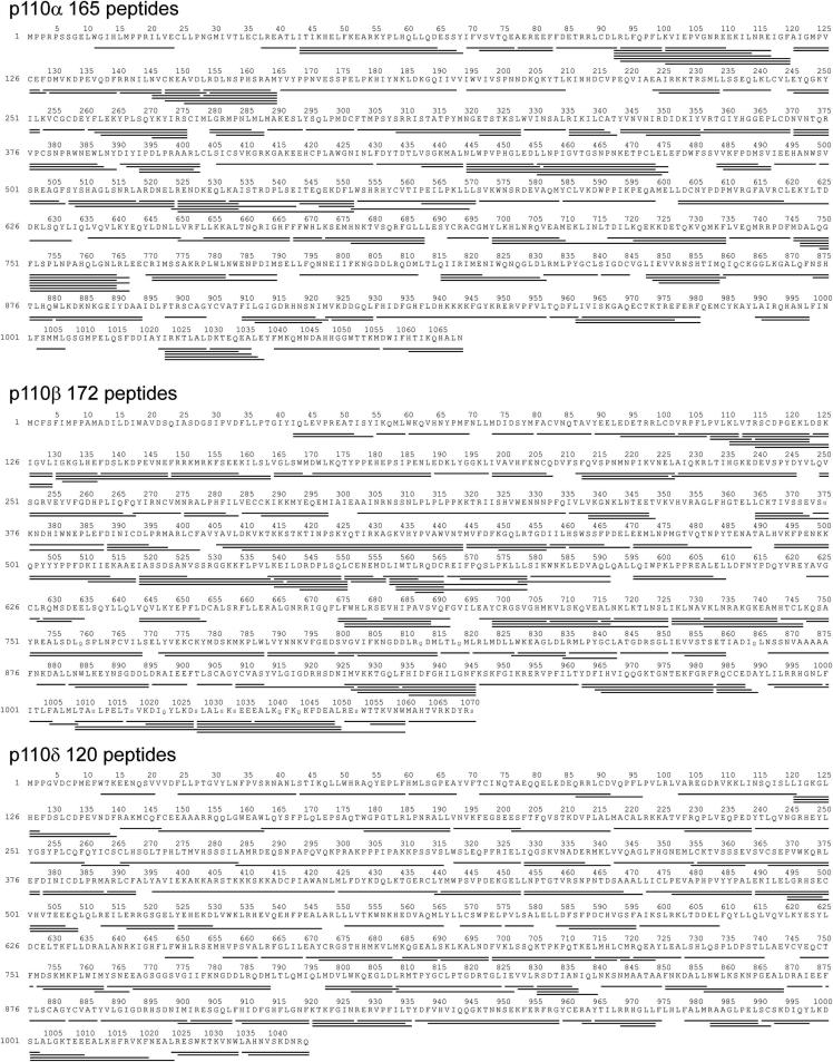
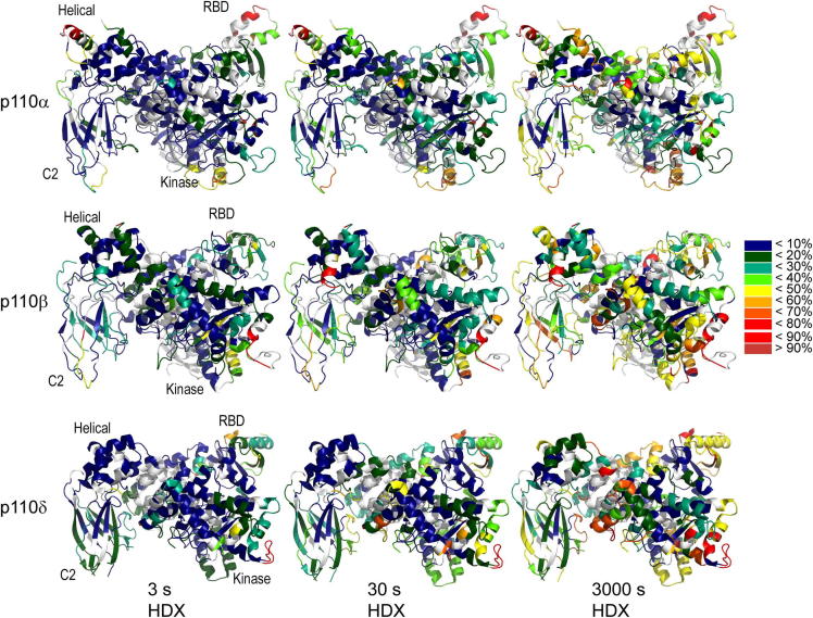
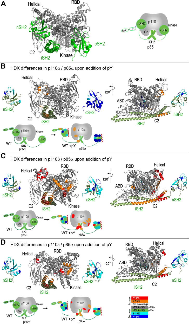
The catalytic subunits of all class IA phosphoinositide 3-kinases (PI3Ks) associate with identical p85-related subunits and phosphorylate PIP2 yielding PIP3, but they can vary greatly in the signaling pathways in which they participate. The binding of the p85 subunit to the p110 catalytic subunits is constitutive, and this inhibits activity, but some of the inhibitory contacts are reversible and subject to regulation. Interaction with phosphotyrosine-containing peptides (RTK-pY) releases a subset of these inhibitory contacts. Hydrogen/deuterium exchange mass spectrometry (HDX-MS) provides a map of the dynamic interactions unique to each of the isotypes. RTK-pY binding exposes the p110 helical domains for all class IA enzymes (due to release of the nSH2 contact) and exposes the C-lobe of the kinase domains of p110β and p110δ (resulting from release of the cSH2 contact). Consistent with this, our in vitro assays show that all class IA isoforms are inhibited by the nSH2, but only p110β and p110δ are inhibited by the cSH2. While a C2/iSH2 inhibitory contact exists in all isoforms, HDX indicates that p110β releases this contact most readily. The unique dynamic relationships of the different p110 isozymes to the p85 subunit may facilitate new strategies for specific inhibitors of the PI3Ks.


Figures

Fig. S1
Fig. S1
Pepsin digested peptide coverage map of p110α, p110β, and p110δ. Identified and analyzed pepsin-digested peptides are shown under the primary sequence of p110 catalytic isoforms. Peptide digestion protocols was optimized for WT full length p110/p85α heterodimers, and allowed for the identification of 165 peptides for p110α, 172 peptides for p110β, and 120 peptides for p110δ.
Fig. S2
Fig. S2
Pepsin digested peptide coverage map of p85α. Identified and analyzed pepsin-digested peptides are shown under the primary sequence of the p85 regulatory subunit. Peptide digestion protocols was optimized for WT full length p110/p85 heterodimers, and allowed for the identification of 107 peptide for p85α.
Fig. S4
Fig. S4
Global H/D levels mapped onto each p110 catalytic isoform. The relative percent deuteration of all peptic peptides shown in Fig. S3 are used to color the structures of the three catalytic isoforms at three timepoints (3HHM, 2Y3A, 2WXH) according to the legend.
Fig. S5
Fig. S5
For the entire set of peptides for p85 (107 peptides), p110α (165 peptides), p110β (172 peptides), and p110δ (120 peptides) the integrated # of deuterium difference in exchange at all four timepoints between each condition labeled was measured. Data represent mean ± SD of two independent experiments. The x axis was generated by taking the central residue of each peptide.
Fig. 1
Fig. 1
PI3K activity assays. A. ATPase activity of all class IA PI3K isoforms in the absence and presence of bis-phosphorylated PDGFR peptide (pY). This assay is based on displacement of the ADP-Alexa 633 tracer from the ADP2 antibody, dependent on the production of ADP. B. Lipid kinase activity assay of all class IA PI3K isoforms with varying amounts of pY (from 0.1 to 100 nM). Assays measured 32P-PIP3 production in the presence of 10 nM enzyme, 100 μM ATP, and 5% PIP2 lipid vesicles at a concentration of 1 mg/ml. The amount of lipid kinase activity for p110α and p110β in the presence of 0.4 nM pY is shown in the bar graph. C. The effect of nSH2 (K379E) and cSH2 (Y685A) mutations on lipid kinase activity. The lipid kinase activity of full-length recombinant p110/p85 complexes containing the indicated p85 mutations are shown in the presence and absence of 50 μM pY. D. The pY sensitivity of lipid kinase activity was determined for all class IA PI3K isoforms in the presence of either the wild type or the Y685A mutant of p85 as described above.
Fig. 2
Fig. 2
Basal hydrogen/deuterium exchange differences in the p85 regulatory subunit in the presence of different p110 catalytic subunits. A. Domain organization of the different p110 catalytic and p85 regulatory subunits. B. Differences in HDX in p85 between the p110α and p110β complex. In the left panel, a structural model of the nSH2, iSH2, and cSH2 of p85 bound to p110β is shown as a reference (a composite image generated from PDB ID 2Y3A, 3HHM, 1H9O and 2V1Y). In the right panel, peptides spanning p85α that showed both greater than 6% and 0.5 Da changes in H/D exchange in between p85 bound to p110α and p110β are colored on the nSH2, iSH2, and cSH2 and mapped onto a model of p85 bound to p110α (generated from PDB ID 3HHM, 1H9O, and 2V1Y). A simplified schematic representation of HDX differences is shown underneath the structural model. C. Differences in HDX in p85 between the p110α and p110δ complex. In the left panel, structural model of the nSH2, iSH2, and cSH2 of p85 bound to p110δ is shown as a reference (generated from PDB ID 2WXH, 3HHM, 1H9O and 2V1Y). In the right panel, peptides spanning p85α that showed both greater than 6% and 0.5 Da changes in H/D exchange in between p85 bound to p110α and p110δ are colored on the nSH2, iSH2, and cSH2 and mapped onto a model of p85 bound to p110α. (For interpretation of the references to color in this figure legend, the reader is referred to the web version of this article.)
Fig. 3
Fig. 3
HDX differences in the p110 catalytic and p85 regulatory subunits in the presence of pY. A. The structural model of SH2 binding to the p110 catalytic subunit based on the crystal structures of p110α bound to the niSH2 fragment of p85, and p110β bound to the icSH2 fragment of p85. The p85 is colored green and the p110 subunit is gray. B. Peptides that showed HDX differences in p110α/p85α in the presence of 15 μM pY are mapped and colored onto a model of the p110α/p85α complex. A simplified schematic representation to the right of the structural model helps orient the location of HDX differences. C. Peptides that showed HDX differences in p110β/p85α in the presence of 15 μM pY are mapped and colored onto a model of the p110β/p85α complex. D. Peptides that showed HDX differences in p110δ/p85α in the presence of 15 μM pY are mapped and colored onto a model of the p110δ/p85α complex. (For interpretation of the references to color in this figure legend, the reader is referred to the web version of this article.)
Fig. 4
Fig. 4
Differential regulation of the class IA PI3Ks by p85. Schematic representation of inhibitory interactions between the p110 and p85 subunits. The p110α subunit has only two inhibitory ‘brakes’ on kinase activity, one from the nSH2 and one from the iSH2. In p110β there are three brakes on lipid kinase activity, due to inhibitory contacts with the nSH2, iSH2, and cSH2, however our HDX results show that the nSH2 and iSH2 ‘brakes’ are slightly disrupted in the basal state compared to p110α. In p110δ there are also three ‘brakes’ on activity from the nSH2, iSH2, and cSH2, however in this case only the nSH2 ‘brake’ is partially disrupted in the basal state, and the iSH2 ‘brake’ remains fully inhibitory.

References

    1. Beeton C.A., Chance E.M., Foukas L.C., Shepherd P.R. Comparison of the kinetic properties of the lipid- and protein-kinase activities of the p110α and p110β catalytic subunits of class-Ia phosphoinositide 3-kinases. Biochem J. 2000;350(2):353–359. - PMC - PubMed
    1. Berndt A., Miller S., Williams O., Le D., Houseman B., Pacold J. The p110δ structure: mechanisms for selectivity and potency of new PI(3)K inhibitors. Nat Chem Biol. 2010;6:117–124. - PMC - PubMed
    1. Breeze A.L., Kara B.V., Barratt D.G., Anderson M., Smith J.C., Luke R.W. Structure of a specific peptide complex of the carboxy-terminal SH2 domain from the p85 subunit of phosphatidylinositol 3-kinase. Embo J. 1996;15:3579–3589. - PMC - PubMed
    1. Brown J., Auger K. Phylogenomics of phosphoinositide lipid kinases: perspectives on the evolution of second messenger signaling and drug discovery. BMC Evol Biol. 2011;11:1–14. - PMC - PubMed
    1. Burke J., Vadas O., Berndt A., Finegan T., Perisic O., Williams R. Dynamics of the phosphoinositide 3-Kinase p110δ interaction with p85α and membranes reveals aspects of regulation distinct from p110α. Structure. 2011;19:1127–1137. - PMC - PubMed

Publication types

MeSH terms

LinkOut - more resources