. 2013 Jan 11;339(6116):227-230.
doi: 10.1126/science.1229663. Epub 2012 Nov 29.

Natively inhibited Trypanosoma brucei cathepsin B structure determined by using an X-ray laser

Affiliations

Natively inhibited Trypanosoma brucei cathepsin B structure determined by using an X-ray laser

Lars Redecke et al. Science. .

Abstract

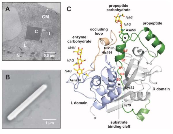
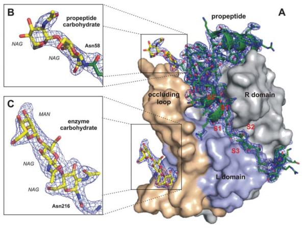
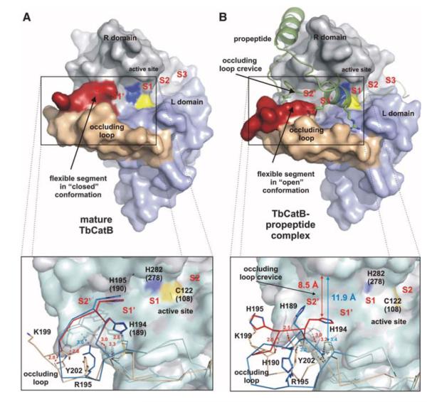
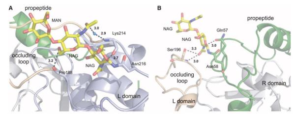
The Trypanosoma brucei cysteine protease cathepsin B (TbCatB), which is involved in host protein degradation, is a promising target to develop new treatments against sleeping sickness, a fatal disease caused by this protozoan parasite. The structure of the mature, active form of TbCatB has so far not provided sufficient information for the design of a safe and specific drug against T. brucei. By combining two recent innovations, in vivo crystallization and serial femtosecond crystallography, we obtained the room-temperature 2.1 angstrom resolution structure of the fully glycosylated precursor complex of TbCatB. The structure reveals the mechanism of native TbCatB inhibition and demonstrates that new biomolecular information can be obtained by the "diffraction-before-destruction" approach of x-ray free-electron lasers from hundreds of thousands of individual microcrystals.


Figures

Fig. 1
Fig. 1
In vivo grown crystals and three-dimensional structure of the TbCatB-propeptide complex. (A) Transmission electron microscopy (EM) of an infected Sf9 insect cell showing a crystal of overexpressed TbCatB inside the rough endoplasmic reticulum that is cut perpendicular to its long axis. N, nucleus; L, lysosome; C, crystal; CM, cell membrane. (B) Scanning EM of a single TbCatB crystal after isolation. (C) Cartoon plot of the TbCatB-propeptide complex exhibiting the typical papain-like fold of cathepsin B–like proteases (supplementary text S1). Gray, R domain; blue, L domain; beige, occluding loop. The native propeptide (green) blocks the active site. The subsites of the substrate-binding cleft N-terminal (nonprime: S2, S3) and C-terminal (prime: S1′, S2′) to the active site (S1) have been identified by comparison with the human CatB structure (13) and labeled (red) according to Schechter and Berger (27). Two N-linked carbohydrate structures (yellow) consist of N-acetylglucosamine (NAG) and mannose (MAN) residues (yellow, carbon atoms; blue, nitrogen atoms; red, oxygen atoms).
Fig. 2
Fig. 2
Quality of the calculated electron density. (A) Surface representation of the TbCatB-propeptide complex solved by molecular replacement using the mature TbCatB structure (11) as a search model. The solution revealed additional electron density (2FobsFcalc, 1σ, blue) of the propeptide (green) that is bound to the V-shaped substrate-binding cleft and of two carbohydrate structures (yellow) N-linked to the propeptide (B) and to the mature enzyme (C). The propeptide, as well as both carbohydrates, are well-defined within the electron density map (blue), which confirms that the phases are not biased by the search model. Color codes correspond to Fig. 1C.
Fig. 3
Fig. 3
Occluding loop conformations of mature and propeptide-inhibited TbCatB. (A) Surface representation of mature TbCatB (11) showing the occluding loop (rigid part, beige; flexible part, red) in the closed conformation. A loop segment blocks the S2′ site and part of the S1′ site of the substrate-binding cleft. (B) Propeptide binding (green) shifts the flexible occluding loop segment (red) into an open conformation, which exposes the entire prime subsite. The insets compare the Tb (gray) and human (cyan) surface representations of mature and propeptide bound CatB. Almost superimposable Cα chains of the human (blue) and Tb (beige/red) occluding loops indicate a conservation of the closed loop conformation in the mature enzymes (A), whereas the open conformations show significant differences (B). Four H bonds maintained during conformational transition restricted the flexible loop segment to four residues (red) in TbCatB, which opens a crevice of ~8.5 Å. In contrast, only one H bond is maintained in the open loop conformation of human procathepsin B (13). Thus, the mobile segment of the human occluding loop (blue) comprises 10 residues and exposes an enlarged occluding loop crevice (supplementary text S5). Residues are labeled according to TbCatB, referring to the corresponding human CatB numbering in parentheses, if applicable.
Fig. 4
Fig. 4
Glycosylation of the TbCatB-propeptide complex. (A) Enzyme carbohydrate structure comprising two NAG and one MAN residue (yellow) N-linked to Asn216 C-terminal of the occluding loop (beige). The carbohydrate structure connects both occluding loop strands by two direct and one water-bridged H bond (black dashed lines). (B) Propeptide glycosylation site comprising two NAG units (yellow) at Asn58 within the kinked region of the propeptide (green). The propeptide carbohydrate structure forms an H bond to Gln57 of the propeptide and two H bonds to Ser196 at the tip of the occluding loop (beige), which stabilize its open conformation. Color codes correspond to Fig. 1C.

Comment in

Similar articles

Cited by

  • Serial femtosecond crystallography of G protein-coupled receptors.
    Liu W, Wacker D, Gati C, Han GW, James D, Wang D, Nelson G, Weierstall U, Katritch V, Barty A, Zatsepin NA, Li D, Messerschmidt M, Boutet S, Williams GJ, Koglin JE, Seibert MM, Wang C, Shah ST, Basu S, Fromme R, Kupitz C, Rendek KN, Grotjohann I, Fromme P, Kirian RA, Beyerlein KR, White TA, Chapman HN, Caffrey M, Spence JC, Stevens RC, Cherezov V. Liu W, et al. Science. 2013 Dec 20;342(6165):1521-4. doi: 10.1126/science.1244142. Science. 2013. PMID: 24357322 Free PMC article.
  • A tool for visualizing protein motions in time-resolved crystallography.
    Wickstrand C, Katona G, Nakane T, Nogly P, Standfuss J, Nango E, Neutze R. Wickstrand C, et al. Struct Dyn. 2020 Apr 1;7(2):024701. doi: 10.1063/1.5126921. eCollection 2020 Mar. Struct Dyn. 2020. PMID: 32266303 Free PMC article.
  • Biophysical highlights from 54 years of macromolecular crystallography.
    Richardson JS, Richardson DC. Richardson JS, et al. Biophys J. 2014 Feb 4;106(3):510-25. doi: 10.1016/j.bpj.2014.01.001. Biophys J. 2014. PMID: 24507592 Free PMC article. Review.
  • Lipidic cubic phase injector facilitates membrane protein serial femtosecond crystallography.
    Weierstall U, James D, Wang C, White TA, Wang D, Liu W, Spence JC, Bruce Doak R, Nelson G, Fromme P, Fromme R, Grotjohann I, Kupitz C, Zatsepin NA, Liu H, Basu S, Wacker D, Han GW, Katritch V, Boutet S, Messerschmidt M, Williams GJ, Koglin JE, Marvin Seibert M, Klinker M, Gati C, Shoeman RL, Barty A, Chapman HN, Kirian RA, Beyerlein KR, Stevens RC, Li D, Shah ST, Howe N, Caffrey M, Cherezov V. Weierstall U, et al. Nat Commun. 2014;5:3309. doi: 10.1038/ncomms4309. Nat Commun. 2014. PMID: 24525480 Free PMC article.
  • Progress and outlook in structural biology of large viral RNAs.
    Cantara WA, Olson ED, Forsyth KM. Cantara WA, et al. Virus Res. 2014 Nov 26;193:24-38. doi: 10.1016/j.virusres.2014.06.007. Epub 2014 Jun 21. Virus Res. 2014. PMID: 24956407 Free PMC article. Review.

References

    1. Frearson JA, et al. Nature. 2010;464:728. - PMC - PubMed
    1. Barrett MP, Boykin DW, Brun R, Tidwell RR. Br. J. Pharmacol. 2007;152:1155. - PMC - PubMed
    1. Alsford S, et al. Nature. 2012;482:232. - PMC - PubMed
    1. Fairlamb AH. Trends Parasitol. 2003;19:488. - PubMed
    1. Bryant C, et al. Bioorg. Med. Chem. Lett. 2009;19:6218. - PMC - PubMed

Publication types

Associated data