Review
. 2012 Dec;11(12):937-57.
doi: 10.1038/nrd3871.

Protein kinase C, an elusive therapeutic target?

Affiliations
Review

Protein kinase C, an elusive therapeutic target?

Daria Mochly-Rosen et al. Nat Rev Drug Discov. 2012 Dec.

Abstract

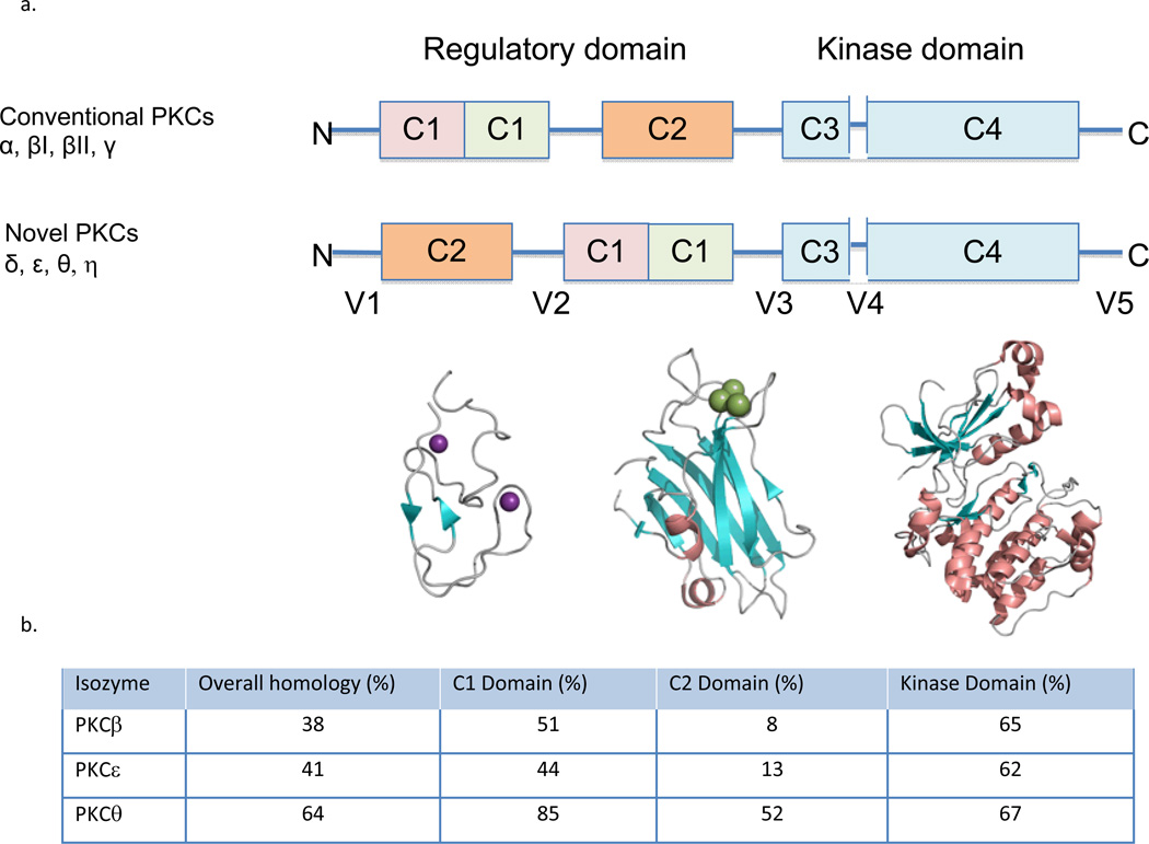
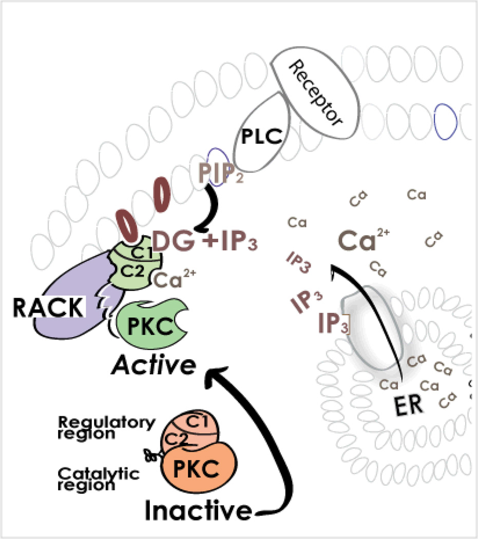
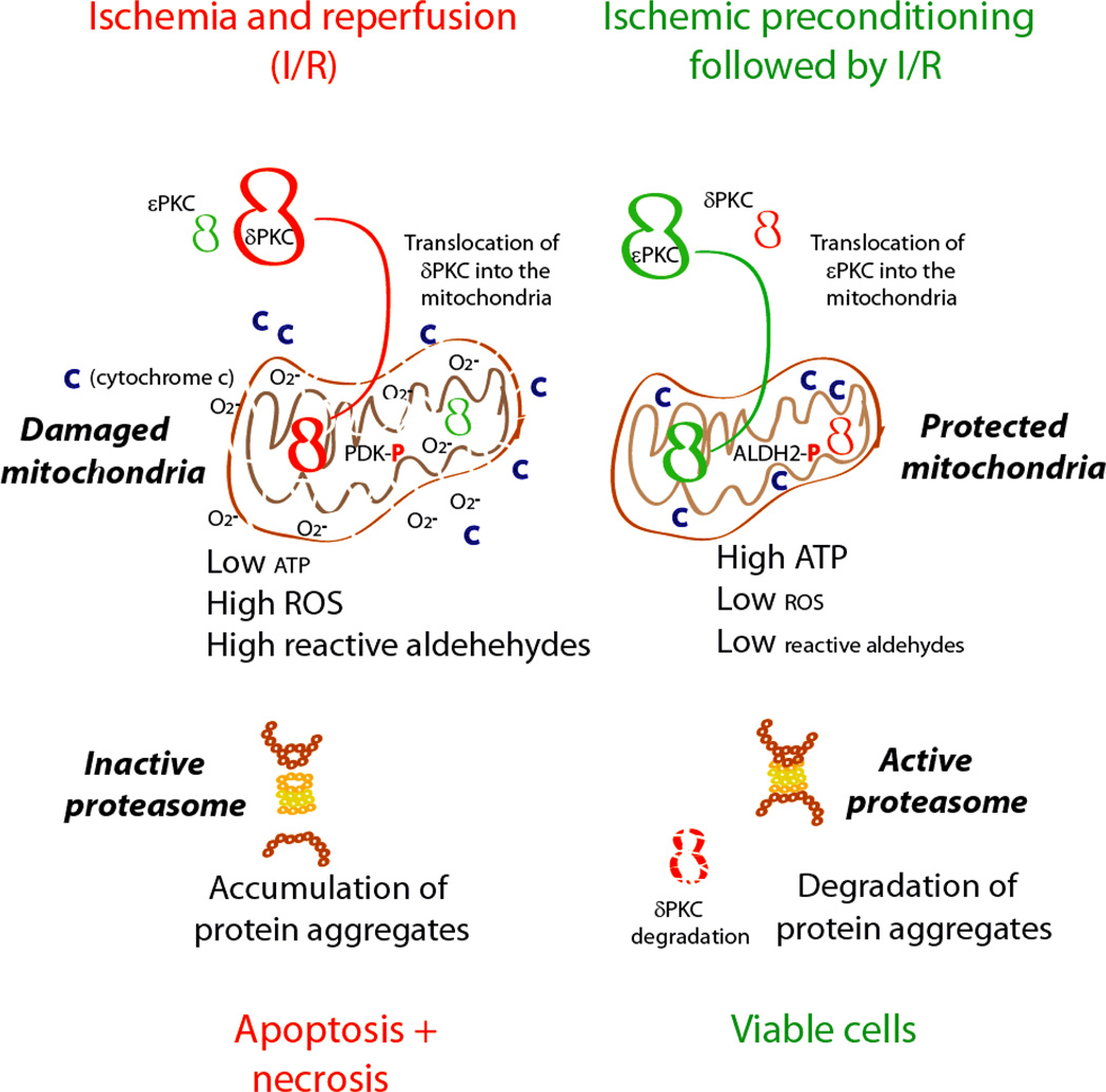
Protein kinase C (PKC) has been a tantalizing target for drug discovery ever since it was first identified as the receptor for the tumour promoter phorbol ester in 1982. Although initial therapeutic efforts focused on cancer, additional indications--including diabetic complications, heart failure, myocardial infarction, pain and bipolar disorder--were targeted as researchers developed a better understanding of the roles of eight conventional and novel PKC isozymes in health and disease. Unfortunately, both academic and pharmaceutical efforts have yet to result in the approval of a single new drug that specifically targets PKC. Why does PKC remain an elusive drug target? This Review provides a short account of some of the efforts, challenges and opportunities in developing PKC modulators to address unmet clinical needs.


Figures

Figure 1
Figure 1
a. Domain composition of PKC family members is shown in a stick scheme (not to scale). The structure of PKCβII (3PFQ) by domains; the diacylglycerol- binding C1a domain, the phosphatidylserine- (and calcium-) binding C2 domain, and the kinase domain. The secondary structures are α helix (in orange), β strands (in cyan) and loops (in gray), Zn2+ in purple and Ca2+ in green. b. The homology of different PKC isozymes to PKCδ. The C2 domain is the least conserved between the isozymes as compared with the other domains, suggesting that pharmacological tools that focus on the C2 domain are more likely to be isozyme-selective.
Figure 2
Figure 2. What processes lead to dissociation of the intra-molecular inhibitory interaction in PKC and to activation of the enzyme?
All PKCs require the binding of diacylglycerol (DG) to the C1 domain of the regulatory region for activation. Conventional PKCs also require calcium (Ca2+) binding to the C2 domain of the regulatory region. These second-messengers are generated following the binding of certain hormones, neurotransmitters or growth factors to their corresponding receptors. The consequent activation of membrane-associated phospholipase C (PLC) results in hydrolysis of phosphatidylinositol-bisphosphate (PIP2), a membrane phospholipid, to DG and inositol trisphosphate (IP3). IP3, in turn, triggers the release of calcium from the endoplasmic reticulum (ER), causing a rise in cytosolic Ca2+ concentration. Therefore, a single event (receptor-dependent phospholipase C activation) leads to generation of the two second-messengers that are required to activate both the conventional and novel PKCs. The rise in DG and Ca2+ leads to activation of PKC and its translocation from the cytosol to the plasma membranes as well as to other subcellular locations, where each isozyme interacts with its anchoring protein, RACK. When bound to RACK and the second messenger activators DG (and Ca2+ for the conventional PKC isozymes), PKC is then active, phosphorylating a number of substrates that are nearby, thus leading to diverse cellular responses.
Figure 3
Figure 3. The role of PKC isozymes in ischemic heart disease
Shown is how prolonged ischemia and reperfusion results in activation of PKCδ more than PKCε, leading to translocation of PKCδ into the mitochondria, phosphorylation of pyruvate dehydrogenase kinase (PDK), which in turn phosphorylates pyruvate dehydrogenase and reduction in TCA cycle and ATP regeneration. Mitochondrial dysfunction leads to higher ROS production and lipid peroxidation leads to accumulation of reactive oxygen species (ROS) and toxic aldehydes, such as 4-hydroxynonenal (4HNE), that interact and inactivate macromolecules including proteins, DNA and lipids. Mitochondrial dysfunction and increase in ROS leads to both apoptosis, and necrosis and severe cardiac dysfunction. In contrast, ischemic preconditioning prior to prolonged ischemia and reperfusion provides cardioprotection by activating more PKCε, which also translocates into the mitochondria and prevents mitochondrial dysfunction induced by prolonged ischemia and reperfusion. PKCε-mediated protection occurs, in part, by PKCε phosphorylation and actvation of aldehyde dehydrogenase 2 (ALDH2). Activated ALDH2 metabolizes aldehydes, such as 4HNE, thus reducing the aldehydic load and the mitochondrial and cellular damage. This results in improved mitochondrial functions and faster recovery of ATP levels. The reduced 4HNE levels also prevent direct inactivation of the peroxisome and thus enable fast removal of aggregated proteins. The active proteasome also selectively degrades activated PKCδ, increasing the balance in favor of the cardiac protective PKCε.
Figure 4
Figure 4. Inhibitors of PKC
Inhibitors of ATP binding to the kinase inhibit the phosphorylation of all the substrates (e.g., S1, S2, S3) of that isozyme, and to a loss of all cellular responses (a). PKC regulators that target the DG-binding site, and act as activators (b) or inhibitors (not shown). The inhibitors can also inhibit anchoring of the enzyme to its RACK that rings the activated isozyme next to its substrates, thus leading to inhibition of all the physiological responses of that isozyme (c). Theoretically, the active, but non-anchored PKC may phosphorylate new substrates. However, this was not observed perhaps because activation in the absence of RACK binding is very transient and/or because the cytosolic phosphatases remove such non-physiological phosphorylations. The same isozyme is localized to several subcellular locations following activation and therefore near a subset of substrates and away from others. Further, there are reports on direct binding of substrates to PKC at sites that are distinct from the phospho-donor/ phospho-acceptor sites. Therefore, inhibitors of protein-protein interactions at a specific subcellular location or with a specific substrate may provide unique inhibitors of the phosphorylation of one substrate and not the others (d). Such separation-of-function inhibitors will have great value, as each PKC isozyme phosphorylates substrates and not all these phosphorylation events contribute to the pathological condition.

References

    1. Castagna M, et al. Direct activation of calcium-activated, phospholipid-dependent protein-kinase by tumor-promoting phorbo esters. J Biol Chem. 1982;257:7847–7851. - PubMed
    1. Geraldes P, King GL. Activation of protein kinase C isoforms and its impact on diabetic complications. Circ Res. 2010;106:1319–1331. - PMC - PubMed
    1. Toton E, Ignatowicz E, Skrzeczkowska K, Rybczynska M. Protein kinase Cepsilon as a cancer marker and target for anticancer therapy. Pharmacol Rep. 2011;63:19–29. - PubMed
    1. Inagaki K, Churchill E, Mochly-Rosen D. Epsilon protein kinase C as a potential therapeutic target for the ischemic heart. Cardiovasc Res. 2006;70:222–230. - PubMed
    1. Ferreira JC, Brum PC, Mochly-Rosen D. BetaIIPKC and epsilonPKC isozymes as potential pharmacological targets in cardiac hypertrophy and heart failure. J Mol Cell Cardiol. 2011;51:479–484. - PMC - PubMed

Publication types