

Mycosis fungoides and Sézary syndrome: clinical, histopathological and immunohistochemical review and update
- PMID: 23197199
- PMCID: PMC3699909
- DOI: 10.1590/s0365-05962012000600001
Mycosis fungoides and Sézary syndrome: clinical, histopathological and immunohistochemical review and update
Abstract
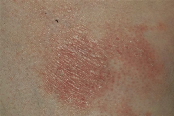
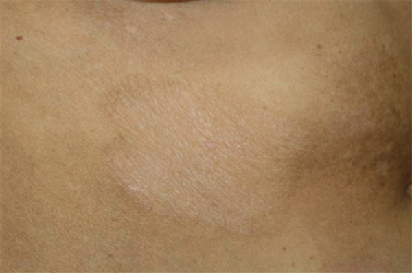
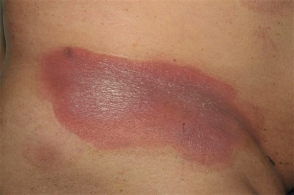
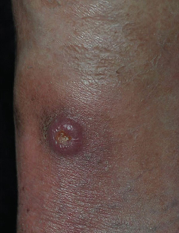
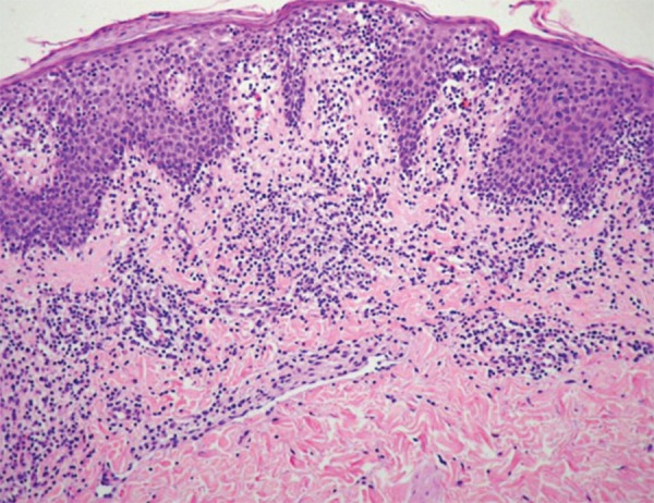
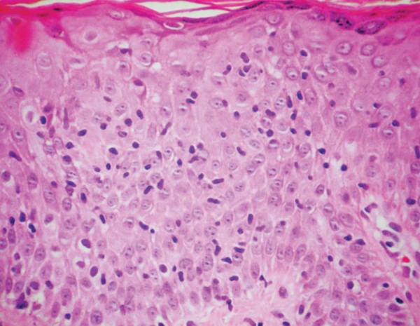
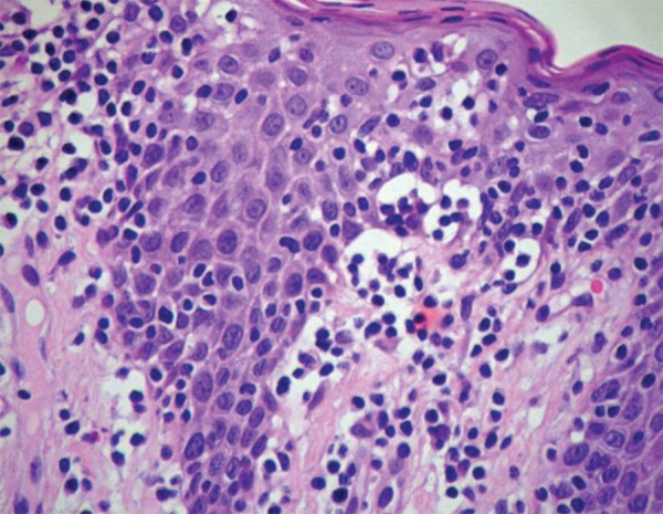
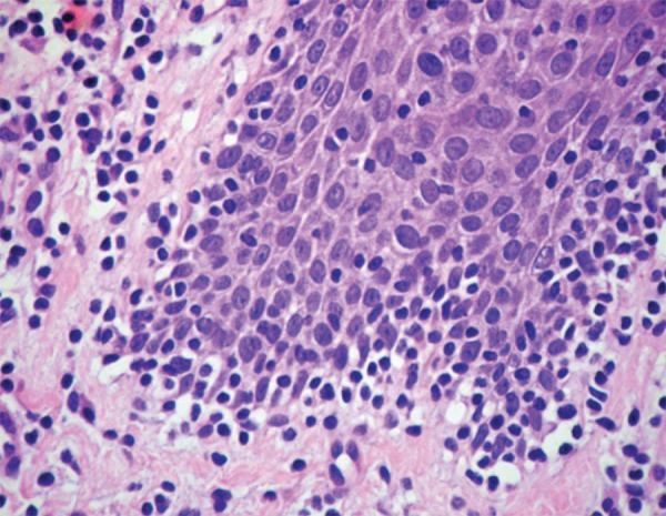
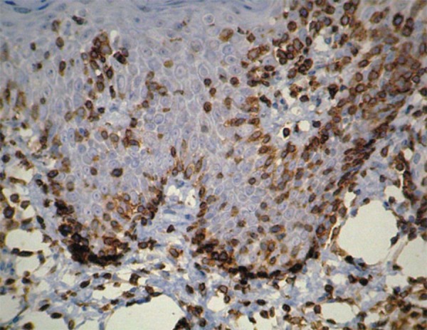
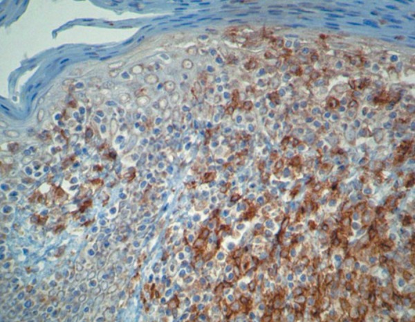
This paper reviews the diagnostic and classificatory concepts of mycosis fungoides and Sézary syndrome in light of the latest normative publications. It describes the great variability of the clinical expression of mycosis fungoides in its early stages as well as the histopathological and immunohistochemical aspects that help with diagnosis. The diagnostic criteria required for characterizing Sézary syndrome and the staging system used for both mycosis fungoides and Sézary syndrome are described.
O artigo revisa os conceitos diagnósticos e de classificação da micose fungóide e da síndrome de Sézary a luz das publicações normativas mais recentes. Descreve a grande variabilidade de expressão clinica da micose fungóide em seus estágios iniciais assim como os aspectos histopatológicos e imuno-histoquímicos auxiliares ao diagnóstico. São descritos os critérios de diagnósticos exigidos para que se caracterize a síndrome de Sézary e o sistema de estadiamento, utilizado para ambas, micose fungóide e síndrome de Sézary.
Conflict of interest statement
Conflict of interest: None
Figures
References
-
- Willemze R, Jaffe ES, Burg G, Cerroni L, Berti E, Swerdlow SH, et al. WHO-EORTC classification for cutaneous lymphomas. Blood. 2005;105:3768–3785. - PubMed
-
- Swerdlow SH, Campo E, Harris NL, Jaffe ES, Pileri SA, Stein H, Thiele J, Vardiman JW, editors. WHO classification of tumors of haematopoietic and lymphoid tissues. Lyon: IARC; 2008.
-
- Rizyl MA, Evens MST, Nelson BP, Rosen ST. T-cell non-Hodgkin lymphoma. Blood. 2008;107:1255–1284. - PubMed
-
- Sanches JA, Jr, Moricz CZM, Neto CF. Processos linfoproliferativos da pele. Parte 2 - Linfomas cutâneos de células T e de células NK. An Bras Dermatol. 2006;81:7–25.
-
- Cerroni L, Gatter K, Kerl H, editors. Skin lymphoma. The illustrated guide. 3rd ed. Oxford, UK: Wiley-Blackwell; 2009. pp. 11–56. Chapter 2, Mycosis fungoides.
Publication types
MeSH terms
LinkOut - more resources
Full Text Sources
Medical