

Gnatophyma--a rare form of rosacea
- PMID: 23197212
- PMCID: PMC3699912
- DOI: 10.1590/s0365-05962012000600014
Gnatophyma--a rare form of rosacea
Abstract
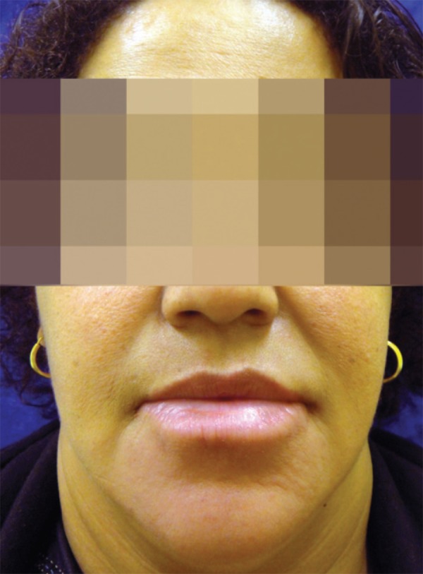
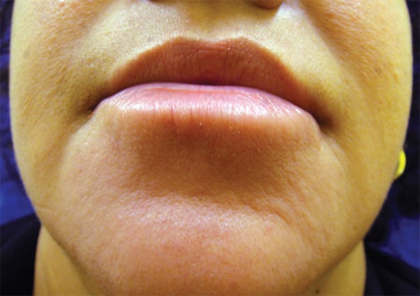
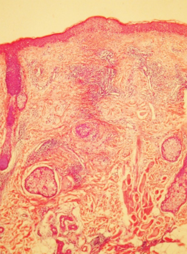
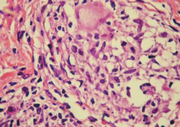
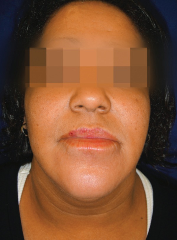
Phyma is the last stage of rosacea and is due to chronic inflammation and edema. It can affect nose (rhinophyma), chin (gnatophyma), forehead (metophyma), ears (otophyma) and eyelids (blepharophyma). Rhinophyma is the most frequent location and there are few reports about gnatophyma. We report the case of a female patient, 41 years old, who had an infiltrated, erythematous, edematous plaque around the chin and lower lip for two years. Histopathology showed perivascular lymphocytic infiltrate, hypertrophied follicles and sebaceous glands, dilated vessels and fibrosis. She was treated with oral tetracycline, oral ivermectin and metronidazole cream with a satisfactory response. The clinical, histopathological and therapeutic response correlation confirmed the diagnosis of gnatophyma, a rare variant of phyma.
Fima é o estágio final da rosácea e ocorre devido ao edema e inflamação crônica. Pode acometer nariz (rinofima), mento (gnatofima), fronte (metofima), orelhas (otofima) e pálpebras (blefarofima). Rinofima é a localização mais encontrada e há raros relatos de gnatofima. Relataremos paciente feminina, 41 anos, que apresentava placa infiltrada, eritêmato-edematosa, em todo o mento e lábio inferior há dois anos. Histopatológico com infiltrado linfocitário perianexial e perivascular, folículos e glândulas sebáceas hipertrofiadas, vasos ectasiados e fibrose perianexial. Foi instituído tratamento com tetraciclina via oral, ivermectina via oral e metronidazol creme com resposta satisfatória. Através da correlação clínica, histopatológica e resposta terapêutica confirmou-se o diagnóstico da variante rara de fima, gnatofima.
Conflict of interest statement
Conflict of interest: None
Figures
Similar articles
-
The Clinical Spectrum of Phyma: A Case Report of Rhino-Metophyma and Review of the Literature.Skin Appendage Disord. 2025 Jun 18. doi: 10.1159/000546890. Online ahead of print. Skin Appendage Disord. 2025. PMID: 40741029 Free PMC article.
-
Gnatophyma and otophyma.J Cutan Med Surg. 2009 Sep-Oct;13(5):266-72. doi: 10.2310/7750.2008.08051. J Cutan Med Surg. 2009. PMID: 19769836
-
Clinical and histological variants of rhinophyma, including nonsurgical treatment modalities.Facial Plast Surg. 1998;14(4):241-53. doi: 10.1055/s-2008-1064456. Facial Plast Surg. 1998. PMID: 11816064 Review.
-
Rhinophyma.2023 Aug 8. In: StatPearls [Internet]. Treasure Island (FL): StatPearls Publishing; 2025 Jan–. 2023 Aug 8. In: StatPearls [Internet]. Treasure Island (FL): StatPearls Publishing; 2025 Jan–. PMID: 31335093 Free Books & Documents.
-
Diagnosis and treatment of rosacea fulminans.Dermatology. 1994;188(4):251-4. doi: 10.1159/000247160. Dermatology. 1994. PMID: 8193395 Review.
Cited by
-
Solitary Plaque on the Chin.Indian Dermatol Online J. 2018 Jan-Feb;9(1):75-76. doi: 10.4103/idoj.IDOJ_110_17. Indian Dermatol Online J. 2018. PMID: 29441311 Free PMC article. No abstract available.
-
Two Cases of Gnatophyma, an Unusual Form of Rosacea.Skin Appendage Disord. 2017 Jan;2(3-4):180-182. doi: 10.1159/000453004. Epub 2016 Dec 8. Skin Appendage Disord. 2017. PMID: 28232929 Free PMC article. No abstract available.
-
[Abscessing swelling of the chin in a young woman].Dermatologie (Heidelb). 2024 Oct;75(10):831-833. doi: 10.1007/s00105-024-05366-z. Epub 2024 Jun 10. Dermatologie (Heidelb). 2024. PMID: 38856792 German. No abstract available.
-
Gnathophyma: Two Cases and Review of the Literature.J Clin Aesthet Dermatol. 2024 Apr;17(4):6-7. J Clin Aesthet Dermatol. 2024. PMID: 38638183 Free PMC article. No abstract available.
-
The Clinical Spectrum of Phyma: A Case Report of Rhino-Metophyma and Review of the Literature.Skin Appendage Disord. 2025 Jun 18. doi: 10.1159/000546890. Online ahead of print. Skin Appendage Disord. 2025. PMID: 40741029 Free PMC article.
References
-
- Crawford GH, Pelle MT, James WD. Rosacea: I. Etiology, pathogenesis, and subtype classification. J Am Acad Dermatol. 2004;51:327–341. - PubMed
-
- Wilkin J, Dahl M, Detmar M, Drake L, Feinstein A, Odom R, Powell F, et al. Standard classification of rosacea: report of the National Rosacea Society Expert Committee on the Classification and Staging of Rosacea. J Am Acad Dermatol. 2002;46:584–587. - PubMed
-
- Rohrich RJ, Griffin JR, Adams WP Jr. Rhinophyma: review and update. Plast Reconstr Surg. 2002;110:860–869. - PubMed
-
- Ilyas EN, Hanson MR, Lawrence N, Green JJ. Gnatophyma. J Am Acad Dermatol. 2006;55:165–166. - PubMed
-
- Vidigal MR, Kakihara CT, Gatti TRSR, Tebcherani AJ, Pires MC. Gnatophyma: a rare variant of phyma. Clin Exp Dermatol. 2008;33:743–744. - PubMed
Publication types
MeSH terms
LinkOut - more resources
Full Text Sources
Medical