

Biochemically and histopathologically comparative review of thiamine's and thiamine pyrophosphate's oxidative stress effects generated with methotrexate in rat liver
- PMID: 23197226
- PMCID: PMC3560789
- DOI: 10.12659/msm.883591
Biochemically and histopathologically comparative review of thiamine's and thiamine pyrophosphate's oxidative stress effects generated with methotrexate in rat liver
Abstract
Background: Oxidative liver injury occurring with methotrexate restricts its use in the desired dose. Therefore, whether or not thiamine and thiamine pyrophosphate, whose antioxidant activity is known, have protective effects on oxidative liver injury generated with methotrexate was comparatively researched in rats using biochemical and histopathological approaches.
Material/methods: Thiamine pyrophosphate+methotrexate, thiamine+methotrexate, and methotrexate were injected intraperitoneally in rats for 7 days. After this period, all animals' livers were excised, killing them with high-dose anesthesia, and histopathologic and biochemical investigations were made.
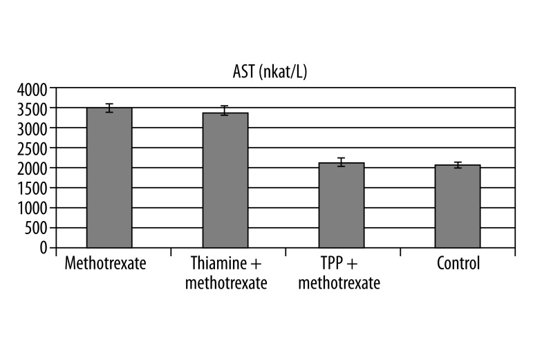
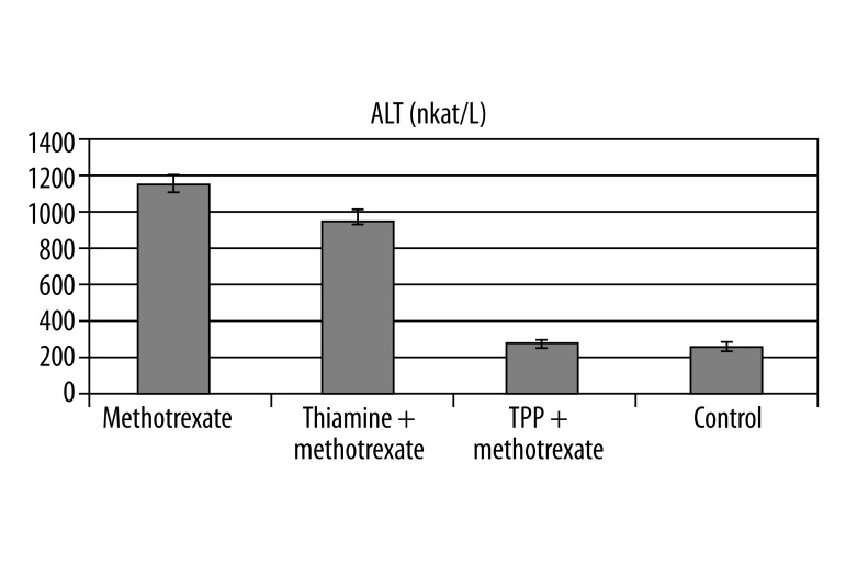
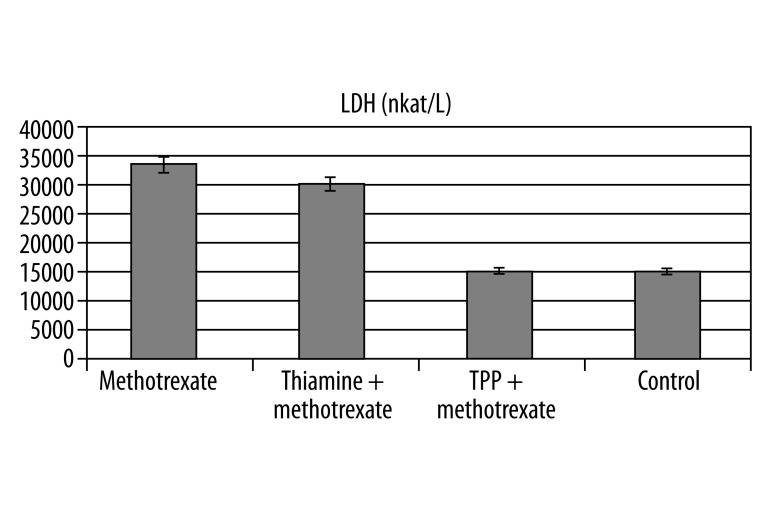
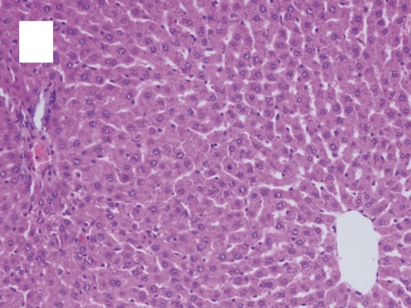
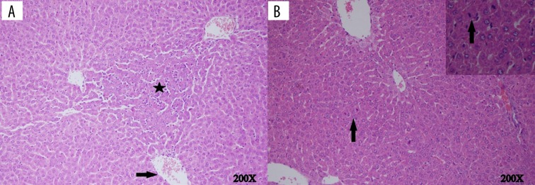
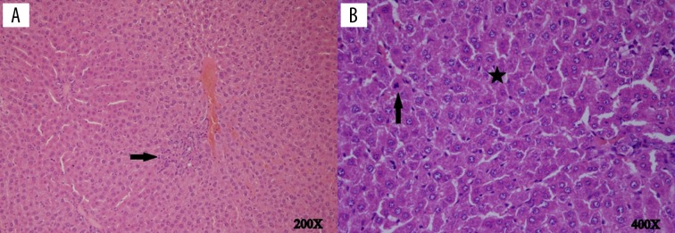
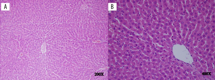
Result: Biochemical results demonstrated a significant elevation in level of oxidant parameters such as MDA and MPO, and a reduction in antioxidant parameters such as GSH and SOD in the liver tissue of the methotrexate group. Also, the quantity of 8-OHdG/dG, a DNA injury product, was higher in the methotrexate group with high oxidant levels and low antioxidant levels, and the quantity of 8-OHdG/dG was in the thiamine pyrophosphate group with low oxidant levels and high antioxidant levels. In the thiamine and control groups, the 8-OHdG/dG rate was 1.48 ± 0.35 pmol/L (P>0.05) and 0.55 ± 0.1 pmol/L (P<0.0001). Thiamine pyrophosphate significantly decreased blood AST, ALT and LDH, but methotrexate and thiamine did not decrease the blood levels of AST, ALT and LDH. Histopathologically, although centrilobular necrosis, apoptotic bodies and inflammation were monitored in the methotrexate group, the findings in the thiamine pyrophosphate group were almost the same as in the control group.
Conclusions: Thiamine pyrophosphate was found to be effective in methotrexate hepatotoxicity, but thiamine was ineffective.
Figures
Similar articles
-
Can thıamıne pyrophosphate prevent desflurane ınduced hepatotoxıcıty ın rats?Acta Cir Bras. 2016 Mar;31(3):168-75. doi: 10.1590/S0102-865020160030000004. Acta Cir Bras. 2016. PMID: 27050787
-
The effects of thiamine and thiamine pyrophosphate on alcohol-induced hepatic damage biomarkers in rats.Eur Rev Med Pharmacol Sci. 2015 Feb;19(4):664-70. Eur Rev Med Pharmacol Sci. 2015. PMID: 25753885
-
The effect of thiamine and thiamine pyrophosphate on oxidative liver damage induced in rats with cisplatin.Biomed Res Int. 2013;2013:783809. doi: 10.1155/2013/783809. Epub 2013 Jun 6. Biomed Res Int. 2013. PMID: 23841092 Free PMC article.
-
Effects of adenosine triphosphate, thiamine pyrophosphate, melatonin, and liv-52 on subacute pyrazinamide proliferation hepatotoxicity in rats.BMC Pharmacol Toxicol. 2025 Mar 24;26(1):67. doi: 10.1186/s40360-025-00901-7. BMC Pharmacol Toxicol. 2025. PMID: 40128909 Free PMC article.
-
Damage induced by paracetamol compared with N-acetylcysteine.J Chin Med Assoc. 2014 Sep;77(9):463-8. doi: 10.1016/j.jcma.2014.01.011. Epub 2014 Jul 12. J Chin Med Assoc. 2014. PMID: 25028290
Cited by
-
The adaptive regulation of thiamine pyrophosphokinase-1 facilitates malignant growth during supplemental thiamine conditions.Oncotarget. 2018 Oct 23;9(83):35422-35438. doi: 10.18632/oncotarget.26259. eCollection 2018 Oct 23. Oncotarget. 2018. PMID: 30459934 Free PMC article.
-
Cytoprotective effects of amifostine, ascorbic acid and N-acetylcysteine against methotrexate-induced hepatotoxicity in rats.World J Gastroenterol. 2014 Aug 7;20(29):10158-65. doi: 10.3748/wjg.v20.i29.10158. World J Gastroenterol. 2014. PMID: 25110444 Free PMC article.
-
Roles of Sulfur Metabolism and Rhodanese in Detoxification and Anti-Oxidative Stress Functions in the Liver: Responses to Radiation Exposure.Med Sci Monit. 2015 Jun 14;21:1721-5. doi: 10.12659/MSM.893234. Med Sci Monit. 2015. PMID: 26071878 Free PMC article. Review.
-
Carvacrol and pomegranate extract in treating methotrexate-induced lung oxidative injury in rats.Med Sci Monit. 2014 Oct 19;20:1983-90. doi: 10.12659/MSM.890972. Med Sci Monit. 2014. PMID: 25326861 Free PMC article.
-
Thiosulfate-Cyanide Sulfurtransferase a Mitochondrial Essential Enzyme: From Cell Metabolism to the Biotechnological Applications.Int J Mol Sci. 2022 Jul 30;23(15):8452. doi: 10.3390/ijms23158452. Int J Mol Sci. 2022. PMID: 35955583 Free PMC article. Review.
References
-
- Cetinkaya A, Bulbuloglu E, Kurutas EB, Kantarceken B. N-acetylcysteine ameliorates methotrexate-induced oxidative liver damage in rats. Med Sci Monit. 2006;12(8):BR274–78. - PubMed
-
- Hytiroglou P, Tobias H, Saxena R, et al. The canals of hering might represent a target of methotrexate hepatic toxicity. Am J Clin Pathol. 2004;121:324–29. - PubMed
-
- Sener G, Eksioglu-Demiralp E, Cetiner M, Ercan F, Yegen BC. Beta-glucan ameliorates methotrexate-induced oxidative organ injury via its antioxidant and immunomodulatory effects. Eur J Pharmacol. 2006;542:170–78. - PubMed
-
- Jolivet J, Cowan KH, Curt GA, Clendeninn NJ, Chabner BA. The pharmacology and clinical use of methotrexate. New Eng J Med. 1983;309:1094–104. - PubMed
-
- Kremer JM, Galivan J, Streckfuss A, Kamen B. Methotrexate metabolism analysis in blood and liver of rheumatoid arthritis patients. Association with hepatic folate deficiency and formation of polyglutamates. Arthritis Rheum. 1986;29:832–35. - PubMed
Publication types
MeSH terms
Substances
LinkOut - more resources
Full Text Sources
Research Materials
Miscellaneous
