

Comparison between normal and loose fragment chondrocytes in proliferation and redifferentiation potential
- PMID: 23197301
- PMCID: PMC3532641
- DOI: 10.1007/s00264-012-1728-x
Comparison between normal and loose fragment chondrocytes in proliferation and redifferentiation potential
Abstract
Purpose: Loose fragments in osteochondritis dissecans (OCD) of the knee require internal fixation. On the other hand, loose fragments derived from spontaneous osteonecrosis of the knee (SONK) are usually removed. However, the difference in healing potential between OCD- and SONK-related loose fragments has not been elucidated. In this study, we investigated proliferative activity and redifferentiation potential of normal cartilage-derived and loose fragment-derived chondrocytes.
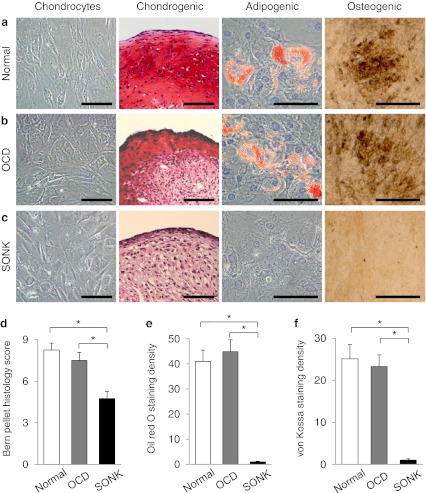
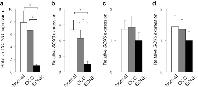
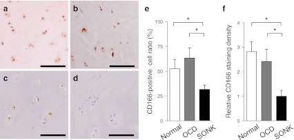
Methods: Cells were prepared from normal articular cartilages and loose fragment cartilages derived from knee OCD and SONK. Cellular proliferation was compared. Redifferentiation ability of pellet-cultured chondrocytes was assessed by real-time PCR analyses. Mesenchymal differentiation potential was investigated by histological analyses. Positive ratio of a stem cell marker CD166 was evaluated in each cartilaginous tissue.
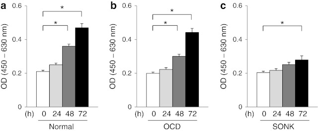
Results: Normal and OCD chondrocytes showed a higher proliferative activity than SONK chondrocytes. Chondrogenic pellets derived from normal and OCD chondrocytes produced a larger amount of safranin O-stained proteoglycans compared with SONK-derived pellets. Expression of chondrogenic marker genes was inferior in SONK pellets. The CD166-positive ratio was higher in normal cartilages and OCD loose fragments than in SONK loose fragments.
Conclusions: The OCD chondrocytes maintained higher proliferative activity and redifferentiation potential compared with SONK chondrocytes. Our results suggest that chondrogenic properties of loose fragment-derived cells and the amount of CD166-positive cells may affect the repair process of osteochondral defects.
Figures
Similar articles
-
Comparison between loose fragment chondrocytes and condyle fibrochondrocytes in cellular proliferation and redifferentiation.J Orthop Sci. 2011 Sep;16(5):589-97. doi: 10.1007/s00776-011-0128-1. Epub 2011 Jul 8. J Orthop Sci. 2011. PMID: 21739103
-
Focal Defects of the Knee Articular Surface: Evidence of a Regenerative Potential Pattern in Osteochondritis Dissecans and Degenerative Lesions.Biomed Res Int. 2017;2017:9036305. doi: 10.1155/2017/9036305. Epub 2017 Jul 9. Biomed Res Int. 2017. PMID: 28770227 Free PMC article.
-
In vitro phenotypic modulation of chondrocytes from knees of patients with osteochondritis dissecans: implications for chondrocyte implantation procedures.J Bone Joint Surg Br. 2012 Jan;94(1):62-7. doi: 10.1302/0301-620X.94B1.27528. J Bone Joint Surg Br. 2012. PMID: 22219249
-
Treatment of unstable knee osteochondritis dissecans in the young adult: results and limitations of surgical strategies-The advantages of allografts to address an osteochondral challenge.Knee Surg Sports Traumatol Arthrosc. 2019 Jun;27(6):1726-1738. doi: 10.1007/s00167-018-5316-5. Epub 2018 Dec 6. Knee Surg Sports Traumatol Arthrosc. 2019. PMID: 30523367 Review.
-
Osteochondritis dissecans knee histology studies have variable findings and theories of etiology.Clin Orthop Relat Res. 2013 Apr;471(4):1127-36. doi: 10.1007/s11999-012-2619-6. Clin Orthop Relat Res. 2013. PMID: 23054514 Free PMC article. Review.
Cited by
-
Single-Cell RNA Sequencing Reveals Transcriptional Changes in the Cartilage of Subchondral Insufficiency Fracture of the Knee.J Inflamm Res. 2022 Nov 5;15:6105-6112. doi: 10.2147/JIR.S385648. eCollection 2022. J Inflamm Res. 2022. PMID: 36386577 Free PMC article.
-
Patient-Specific iPSC-Derived Models Link Aberrant Endoplasmic Reticulum Stress Sensing and Response to Juvenile Osteochondritis Dissecans Etiology.Stem Cells Transl Med. 2023 May 15;12(5):293-306. doi: 10.1093/stcltm/szad018. Stem Cells Transl Med. 2023. PMID: 37184892 Free PMC article.
-
Articular Cartilage Repair of the Knee in Children and Adolescents.Orthop J Sports Med. 2018 Mar 13;6(3):2325967118760190. doi: 10.1177/2325967118760190. eCollection 2018 Mar. Orthop J Sports Med. 2018. PMID: 29568785 Free PMC article. Review.
-
Postoperative change in medial meniscal length in concurrent all-inside meniscus repair with anterior cruciate ligament reconstruction.Int Orthop. 2014 Jul;38(7):1393-9. doi: 10.1007/s00264-013-2238-1. Epub 2013 Dec 18. Int Orthop. 2014. PMID: 24346514 Free PMC article.
-
Postoperative change in the length and extrusion of the medial meniscus after anterior cruciate ligament reconstruction.Int Orthop. 2015 Dec;39(12):2481-7. doi: 10.1007/s00264-015-2704-z. Epub 2015 Feb 19. Int Orthop. 2015. PMID: 25693884
References
-
- Bradley J, Dandy DJ. Osteochondritis dissecans and other lesions of the femoral condyles. J Bone Joint Surg Br. 1989;71:518–522. - PubMed
Publication types
MeSH terms
Substances
LinkOut - more resources
Full Text Sources
Medical
Research Materials
Miscellaneous
