. 2013 Feb;109(4):1097-106.
doi: 10.1152/jn.00304.2012. Epub 2012 Nov 28.

Corticospinal excitability is enhanced after visuomotor adaptation and depends on learning rather than performance or error

Affiliations

Corticospinal excitability is enhanced after visuomotor adaptation and depends on learning rather than performance or error

Hamid F Bagce et al. J Neurophysiol. 2013 Feb.

Abstract

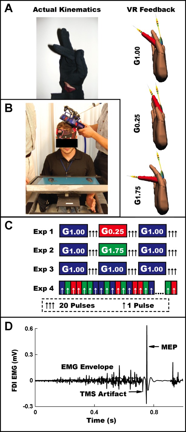
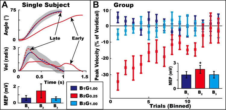
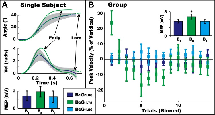
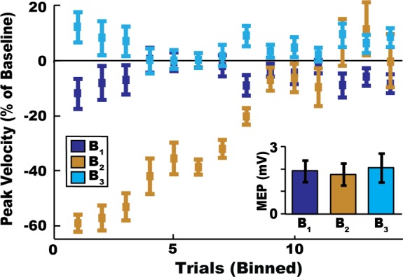
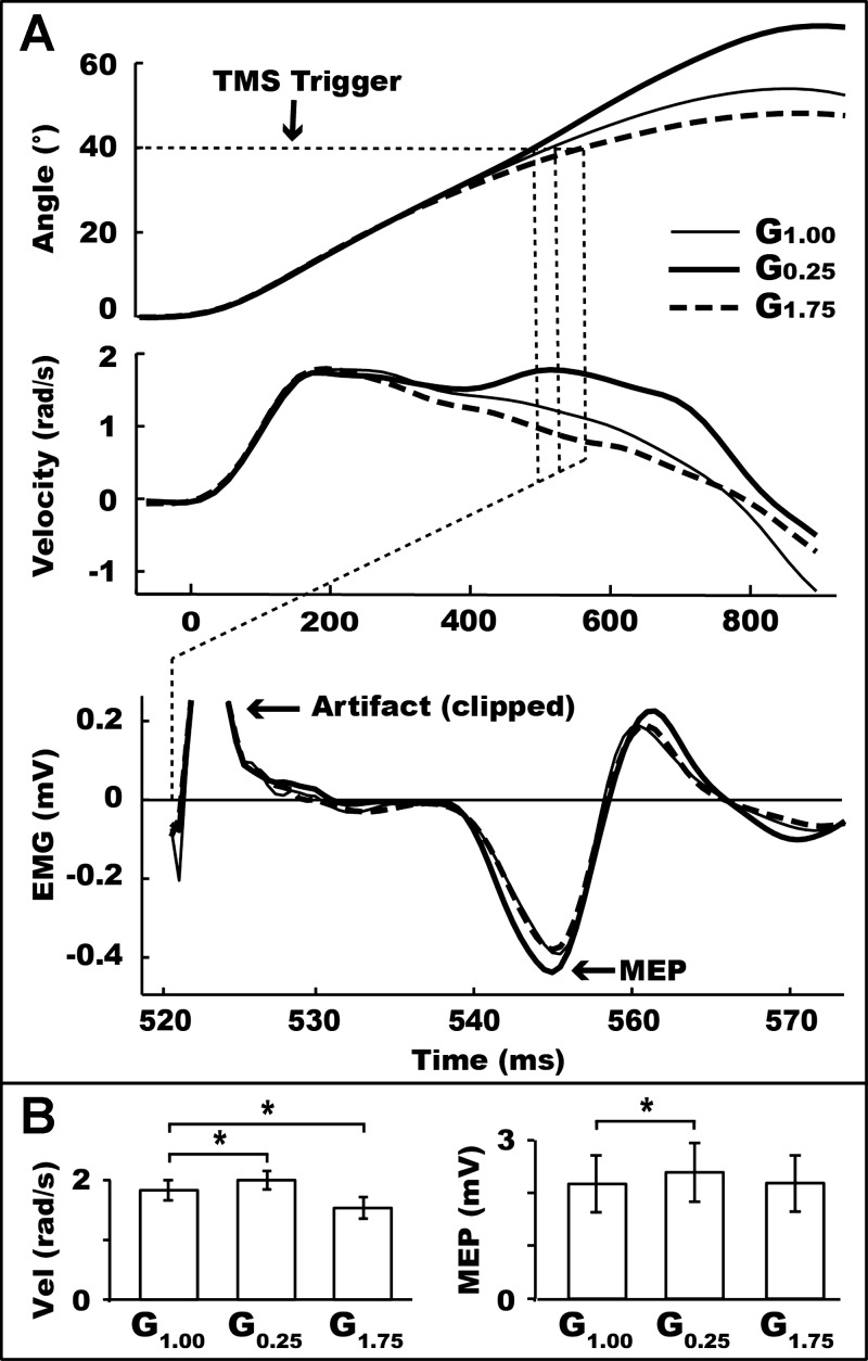
We used adaptation to high and low gains in a virtual reality setup of the hand to test competing hypotheses about the excitability changes that accompany motor learning. Excitability was assayed through changes in amplitude of motor evoked potentials (MEPs) in relevant hand muscles elicited with single-pulse transcranial magnetic stimulation (TMS). One hypothesis is that MEPs will either increase or decrease, directly reflecting the effect of low or high gain on motor output. The alternative hypothesis is that MEP changes are not sign dependent but rather serve as a marker of visuomotor learning, independent of performance or visual-to-motor mismatch (i.e., error). Subjects were required to make flexion movements of a virtual forefinger to visual targets. A gain of 1 meant that the excursions of their real finger and virtual finger matched. A gain of 0.25 ("low gain") indicated a 75% reduction in visual versus real finger displacement, a gain of 1.75 ("high gain") the opposite. MEP increases (>40%) were noted in the tonically activated task-relevant agonist muscle for both high- and low-gain perturbations after adaptation reached asymptote with kinematics matched to veridical levels. Conversely, only small changes in excitability occurred in a control task of pseudorandom gains that required adjustments to large errors but in which learning could not accumulate. We conclude that changes in corticospinal excitability are related to learning rather than performance or error.


Figures

Fig. 1.
Fig. 1.
A: actual finger position (left) and virtual finger position (right) for a single 45° physical angle. Physical angle (performance) was identical and virtual angle (feedback) was augmented (in real time) for each condition within each experiment. Virtual target arrows (for experiments 1, 2, and 3) and virtual finger color change (experiment 4) are shown. VR, virtual reality. B: virtual reality setup. C: condition and trial structure for each experiment. Motor evoked potential (MEP) acquisition is indicated by arrows either after a block of movements (experiments 1, 2, 3) or during a given trial (experiment 4). D: raw and filtered/rectified (thin line) EMG signal acquired from a typical subject in experiment 4. FDI, first dorsal interosseous; TMS, transcranial magnetic stimulation.
Fig. 2.
Fig. 2.
Kinematic and electrophysiological data in experiment 1. A (subject): blue line and shaded region represent the mean (±SD) metacarpophalangeal (MCP) flexion angle (top) and peak angular velocity (middle) for a typical subject, averaged across B1G1.00 and B3G1.00. Mean traces of the first 3 (early, solid red) and last 3 (late, dashed red) trials of B2G0.25 are superimposed. Mean (±SD) MEP bar plot (bottom) for this subject demonstrates increased M1 facilitation immediately after B2G0.25 relative to B1G1.00 and B3G1.00. B (group): group mean (± SE) peak velocity as % change relative to the average veridical trials for each angle for each block. Trials for all 3 target angles are binned together; thus the 42 total trials are represented by 14 bins on the x-axis. Subjects adapted to B2G0.25 by normalizing movement velocity to the level observed in the veridical blocks. Also shown (inset) is group mean (±SE) MEP.
Fig. 3.
Fig. 3.
Subject (A) and group (B) kinematic and electrophysiological data in experiment 2. Labels are as in Fig. 2.
Fig. 4.
Fig. 4.
Kinematic and electrophysiological data in control experiment 3 (see materials and methods). Labels are as in Fig. 2B. Gold color shows the incrementally increasing angular velocity over the course of the block, without any requirement for visuomotor adaptation (as was necessary in experiments 1 and 2). Note the absence of any modulation of M1 excitability at the group level (inset).
Fig. 5.
Fig. 5.
Kinematic and electrophysiological data in experiment 4. A (subject): mean MCP flexion angle (top) and angular velocity (middle) for G1.00, G0.25, and G1.75 trials. Bottom: the same subject's mean MEP traces, which have been realigned in time according to when TMS was triggered. Dotted line marks the time at which the subject attained a 40° flexion angle, resulting in a trigger to signal the TMS pulse. B (group): mean (±SE) instantaneous angular velocity (left) and MEP (right). Asterisk denotes significant effects in a 1-sample paired t-test. To rule out velocity-based confounds on MEPs, data were reanalyzed by excluding G0.25 trials in which angular velocity exceeded 1 SD of the global mean velocity and, similarly, excluding G1.75 trials in which velocity was below 1 SD of the global mean. Blinded reanalysis of MEP data after velocity equalization revealed that MEPs were unaffected.

References

    1. Adkins DL, Boychuk J, Remple MS, Kleim JA. Motor training induces experience-specific patterns of plasticity across motor cortex and spinal cord. J Appl Physiol 101: 1776–1782, 2006 - PubMed
    1. Agostino R, Iezzi E, Dinapoli L, Gilio F, Conte A, Mari F, Berardellli A. Effects of 5 Hz subthreshold magnetic stimulation of primary motor cortex on fast finger movements in normal subjects. Exp Brain Res 180: 105–111, 2007 - PubMed
    1. Aranyi Z, Mathis J, Hess CW, Rosler KM. Task-dependent facilitation of motor evoked potentials during dynamic and steady muscle contractions. Muscle Nerve 21: 1309–1316, 1998 - PubMed
    1. Bindman LJ, Lippold OC, Redfearn JW. The action of brief polarizing currents on the cerebral cortex of the rat (1) during current flow and (2) in the production of long-lasting after-effects. J Physiol 172: 369–382, 1964 - PMC - PubMed
    1. Castro-Alamancos MA, Connors BW. Distinct forms of short-term plasticity at excitatory synapses of hippocampus and neocortex. Proc Natl Acad Sci USA 94: 4161–4166, 1997 - PMC - PubMed

Publication types

LinkOut - more resources