. 2013 Feb;109(4):932-9.
doi: 10.1152/jn.00459.2012. Epub 2012 Nov 28.

Short-term plasticity shapes activity pattern-dependent striato-pallidal synaptic transmission

Affiliations

Short-term plasticity shapes activity pattern-dependent striato-pallidal synaptic transmission

Juhyon Kim et al. J Neurophysiol. 2013 Feb.

Abstract

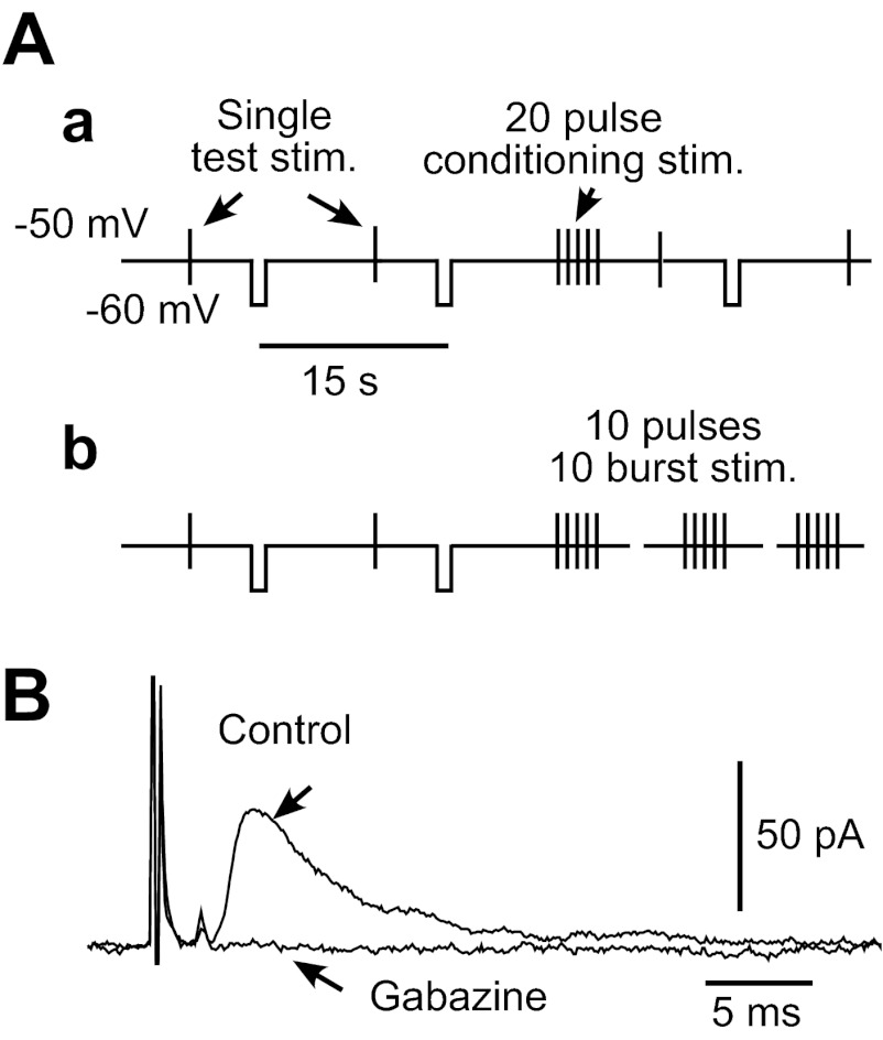
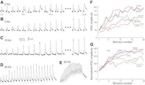
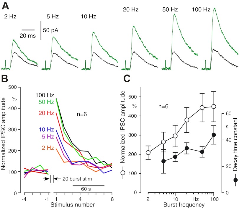
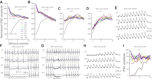
The cortico-striato (Str)-globus pallidus external segment (GPe) projection plays major roles in the control of neuronal activity in the basal ganglia under both normal and pathological conditions. The present study used rat brain-slice preparations to address our hypothesis that the gain of this disynaptic projection is dynamically controlled by activations of short-term plasticity mechanisms of Str-GPe synapses. The Str-GPe projection neurons fire with very different frequency and firing patterns in vivo depending on the condition of the animal. The results show that the Str-GPe synapses have very strong short-term enhancement mechanisms and that repetitive burst activation of the Str-GPe synapses, which mimic oscillatory burst firing of Str neurons, can sustain enhanced states of synaptic transmission for tens of seconds. The results reveal that the short-term enhancement of Str-GPe synapses contributes to the generation of pauses in the firing of GPe neurons and that signal transfer function in the Str-GPe projection is highly dependent on the firing pattern of Str neurons.


Figures

Fig. 1.
Fig. 1.
A: diagrams show stimulus (stim.) and the membrane-clamp schedules used in this study. Globus pallidus external segment (GPe) neurons were voltage clamped at −50 mV to block autonomous firing. Single test stimuli were given every 15 s, and −10 mV pulses were applied 5 s following each test stimulation to monitor the input impedance of the neuron. We used 2 repetitive stimulation schedules: single burst of 20 repetitive pulses with frequencies of 2–100 Hz (a) or 10 bursts of 10 repetitive pulses with 20 or 50 Hz and various interburst intervals (IBIs; b). B: stimulus intensity was adjusted to evoke 30–50 pA inhibitory postsynaptic currents (IPSCs) in test stimulation unless noted otherwise. For all recordings, artificial cerebrospinal fluid contained 10 μM 1,2,3,4-tetrahydro-6-nitro-2,3-dioxo-benzo[f]quinoxaline-7-sulfonamide and 30 μM 3-(2-carboxypiperzin-4-yl)-pro-pyl-1-phosponic acid to block AMPA/kainate and N-methyl-d-aspartate responses. Application of 10 μM gabazine completely abolished the fast IPSCs.
Fig. 2.
Fig. 2.
Responses of GPe neurons to burst striatal (Str) stimulation at various frequencies. A–E: IPSCs recorded from a GPe neuron, voltage clamped at −50 mV. F: plots of IPSC amplitudes shown in A–E. G: group plots of 6 neurons. IPSC amplitudes were normalized with the mean amplitude of 4 IPSCs evoked before burst stimulation.
Fig. 3.
Fig. 3.
Postconditioning enhancement of Str-GPe IPSCs. A: examples of the enhancement of IPSCs. An overlay plot of IPSCs recorded before and 5 s after a conditioning stimulation consisting of 20 pulses with different frequencies. B: group data from 6 neurons show that the degree of enhancement is dependent on the frequency of the conditioning stimuli. The amplitudes of IPSCs were normalized by the mean of 4 IPSCs, evoked before a conditioning stimulation. C: plots of the mean amplitude of the 1st IPSCs evoked after the conditioning and the decay time constant. The decay time constants were estimated from responses to test stimuli applied every 15 s.
Fig. 4.
Fig. 4.
Repetitive burst activation enhanced Str-GPe synapses. A–D: group data show changes in IPSC amplitude recorded from GPe neurons during Str burst stimuli, 10 sets of 10 pulses with different frequencies and IBIs. For each neuron, the amplitudes of the IPSCs were normalized by the average of 4 IPSCs evoked by 4 single stimuli given prior to burst stimuli in 15-s intervals. A and B: IPSCs of GPe neurons to 20 and 50 Hz burst stimulation with 2-s IBIs. The IPSCs to 1st burst stimuli were greatly enhanced (brown lines). The IPSCs to succeeding burst stimuli were started from an enhanced amplitude and declined during burst, with the degree of enhancement and decline depending on the frequency of the stimuli. B–D: compare results of 50 Hz burst stimulation with 2- to 40-s IBIs. The carryover of enhanced IPSCs is clearly visible with 10-s IBIs but is very small with 40-s IBIs. E: responses of a GPe neuron to 50 Hz burst stimulation with 10-s IBIs. F and G: examples of current clamp recordings to show that repetitive burst activation of Str can induce pauses of firing in GPe neurons that were maintained to fire at ∼20 Hz by current injection. Action potentials were truncated, and horizontal lines mark −40 mV. H and I: responses of a GPe neuron to a threshold intensity 50 Hz burst stimulation with 2-s IBIs. The reduction rate of IPSC amplitude during the burst was much lower compared with that of responses to stronger burst stimulation (compare B with I). It is also clear that the threshold intensity at a single stimulus can evoke large IPSCs in burst activation conditions.
Fig. 5.
Fig. 5.
The dopamine D2 receptor agonist quinpirole (10 μM) and lowering extracellular Ca2+ from 2.4 to 1.2 mM reduced IPSC amplitude and slowed development of repetitive stimuli-induced enhancement. A and B: examples of responses of a GPe neuron show enhancement of IPSCs to 20 Hz, 20 pulse burst stimuli in the control and in quinpirole. C: group data compare the development of enhancement before and after quinpirole. The exponential regression curves were computed to compare the progress of the enhancement. The dimension of the time constants (τ) is the stimulus interval. D–F: lowering extracellular Ca2+ from 2.4 to 1.2 mM resulted in similar effects to quinpirole.

References

    1. Baufreton J, Bevan MD. D2-like dopamine receptor-mediated modulation of activity-dependent plasticity at GABAergic synapses in the subthalamic nucleus. J Physiol 586: 2121–2142, 2008 - PMC - PubMed
    1. Catterall WA, Few AP. Calcium channel regulation and presynaptic plasticity. Neuron 59: 882–901, 2008 - PubMed
    1. Chan CS, Shigemoto R, Mercer JN, Surmeier DJ. HCN2 and HCN1 channels govern the regularity of autonomous pacemaking and synaptic resetting in globus pallidus neurons. J Neurosci 24: 9921–9932, 2004 - PMC - PubMed
    1. Chen MT, Morales M, Woodward DJ, Hoffer BJ, Janak PH. In vivo extracellular recording of striatal neurons in the awake rat following unilateral 6-hydroxydopamine lesions. Exp Neurol 171: 72–83, 2001 - PubMed
    1. Connelly WM, Schulz JM, Lees G, Reynolds JN. Differential short-term plasticity at convergent inhibitory synapses to the substantia nigra pars reticulata. J Neurosci 30: 14854–14861, 2010 - PMC - PubMed

Publication types

LinkOut - more resources