. 2012 Nov 30;338(6111):1177-83.
doi: 10.1126/science.1227268.

Three-dimensional structures self-assembled from DNA bricks

Affiliations

Three-dimensional structures self-assembled from DNA bricks

Yonggang Ke et al. Science. .

Abstract

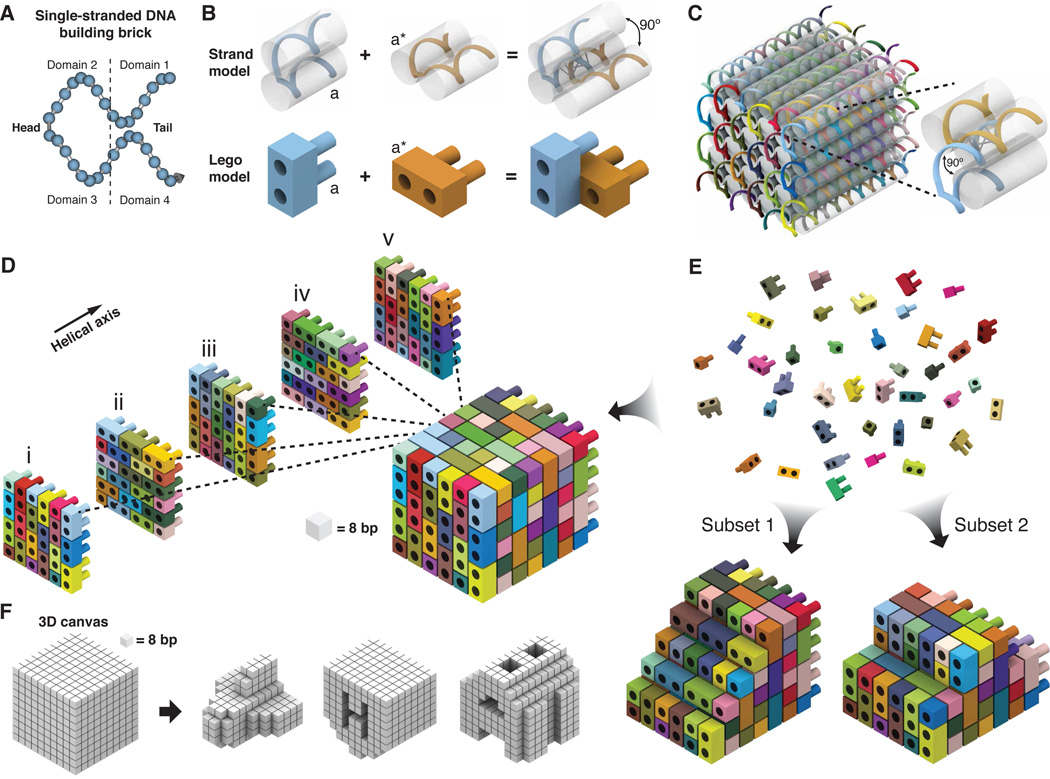
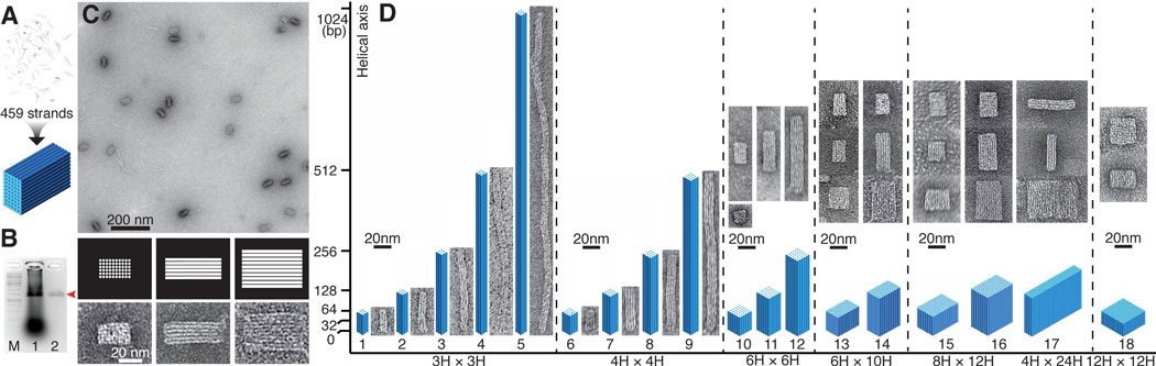
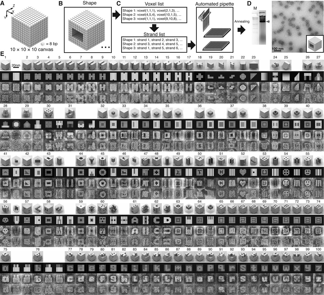
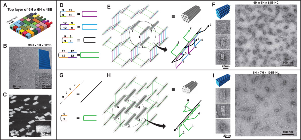
We describe a simple and robust method to construct complex three-dimensional (3D) structures by using short synthetic DNA strands that we call "DNA bricks." In one-step annealing reactions, bricks with hundreds of distinct sequences self-assemble into prescribed 3D shapes. Each 32-nucleotide brick is a modular component; it binds to four local neighbors and can be removed or added independently. Each 8-base pair interaction between bricks defines a voxel with dimensions of 2.5 by 2.5 by 2.7 nanometers, and a master brick collection defines a "molecular canvas" with dimensions of 10 by 10 by 10 voxels. By selecting subsets of bricks from this canvas, we constructed a panel of 102 distinct shapes exhibiting sophisticated surface features, as well as intricate interior cavities and tunnels.


Figures

Fig. 1
Fig. 1
Design of DNA brick structures. (A) A 32-nucleotide four-domain single-stranded DNA brick. Each domain is 8 nucleotides in length. The connected domains 2 and 3 are “head” domains; domains 1 and 4 are “tail” domains. (B) Each two-brick assembly forms a 90° dihedral angle via hybridization of two complementary 8-nucleotide-domains “a” and “a∗”. (C) A molecular model that shows the helical structure of a 6H × 6H × 48B cuboid 3D DNA structure. Each strand has a unique sequence, as indicated by a distinct color. The inset shows a pair of bricks. (D) A Lego-like model of the 6H × 6H × 48B cuboid. Each brick has a unique sequence. The color use is consistent with (B). Half-bricks are present on the boundary of each layer. (E) The 6H × 6H × 48B cuboid is self-assembled from DNA bricks. The bricks are not interchangeable during self-assembly because of the distinct sequence of each brick. Using the 6H × 6H × 48B as a 3D “molecular canvas”, a smaller shape can be designed using a subset of the bricks. (F) 3D shapes designed from a 10 × 10 × 10 voxel “3D canvas”; each voxel fits 8 base-pairs (2.5 nm × 2.5 nm × 2.7 nm).
Fig. 2
Fig. 2
Cuboid structures self-assembled from DNA bricks. (A) DNA bricks self-assembled into a 6H × 10H ×128B cuboid in a one-step thermal annealing process. (B) Agarose gel electrophoresis showing 50% purification recovery efficiency of the 6H × 10H × 128B cuboid. Lane M contains the 1 kb ladder. Lanes 1 and 2 contain unpurified and purified 6H × 10H × 128B cuboid structures, respectively. The red arrow points to the cuboid product band. (C) TEM images of gel purified 6H × 10H × 128B cuboid. Zoomed-in images (bottom) and corresponding computer-generated graphics (middle) show three different projection views. (D) Designs and TEM images of 18 cuboids of a variety of dimensions. Horizontal axis is labeled with the cross-section dimensions of the cuboids; vertical axis is labeled with the lengths of the constituent helices. The lengths are 48B (shape 18), 64B (shapes 1, 6, 10, 13, 15), 120B (shapes 16,17), 128B (shapes 2, 7, 11, 14), 256B (shapes 3, 8, 12), 512B (shapes 4, 9), and 1,024B (shape 5). Each 3D cylinder model is drawn proportionally to the relative dimensions of the cuboid; corresponding TEM images are shown on the right or above.
Fig. 3
Fig. 3
Shapes made from a 3D “molecular canvas”. (A) A 10 × 10 × 10 voxel 3D canvas. Z-axis is the helical axis. Each voxel (8 base-pairs) measures 2.5 nm × 2.5 nm × 2.7 nm. (B) Shapes are designed by editing voxels using 3D modeling software. (C) A computer program recognizes the voxel composition of each shape and generates a list of strands to form this shape. The list then is used to direct an automated liquid-handling robot to mix the strands. (D) After annealing, the shapes are characterized by agarose gel electrophoresis and TEM imaging. Lane M contains the 1 kb ladder. The product band is indicated by the red arrow. (E) Computer-generated models and TEM images of shapes. The top row for each shape depicts a 3D model, followed by a computer-generated projection view, an image averaged from 6 different particles visualized using TEM, and a representative raw TEM image. More raw images are shown in figs. S38–54. In a number of cases, multiple projections are presented. Some shapes with cavities or tunnels are depicted with additional transparent 3D views that highlight the deleted voxels (colored dark gray). For example, the top-right model of shape 32 shows the enclosed cuboid cavity.
Fig. 4
Fig. 4
Generality of DNA brick self-assembly. (A–C) depict the design and construction of a single-layer brick structure. (A) DNA bricks of the top layer of the 6H × 6H × 48B cuboid in Fig. 1D, with the crossovers to the layer below removed. (B) TEM images of a 30H × 1H × 126B rectangle. Top-right inset shows the model of the design. Bottom-right inset contains a zoomed-in image of the structure. (C) AFM images of the 30H × 1H × 126B rectangle. Inset contains a zoomed-in image of the structure. (D–I) depict the designs and constructions of 3D honeycomb-lattice (D–F) and hexagonal-lattice (G–I) brick structures. (D, G) The strands used for honeycomb-lattice (D) and hexagonal-lattice (G) self-assembly. Numbers in the left panels indicate the number of nucleotides in a domain. (E, H) Strand diagrams of an 84-base-pair honeycomb-lattice structure (E) and a 54-base-pair hexagonal-lattice structure (H). The right-bottom image depicts an enlarged image of the circled helix bundle. Strand colors match those described in the right side of (D) or (G). Numbers indicate DNA helices. Note helix 1 and helix 5 are not directly connected in (H). The 9-base half-bricks were merged with their neighboring bricks to form longer strands. (F, I) TEM images of a 6H × 6H × 84B-HC hexagonal-lattice structure (F) and a 6H × 7H × 108B-HL 3D hexagonal-lattice structure (I). 3D model and zoomed-in images of different projection views are shown on the left.

Comment in

References

    1. Seeman NC. Nucleic acid junctions and lattices. J. Theor. Biol. 1982;99:237. - PubMed
    1. Chen J, Seeman NC. Synthesis from DNA of a molecule with the connectivity of a cube. Nature. 1991;350:631. - PubMed
    1. Fu TJ, Seeman NC. DNA double-crossover molecules. Biochemistry. 1993;32:3211. - PubMed
    1. Winfree E, Liu F, Wenzler LA, Seeman NC. Design and self-assembly of two-dimensional DNA crystals. Nature. 1998;394:539. - PubMed
    1. Yurke B, Turberfield AJ, Mills AP, Jr, Simmel FC, Neumann JL. A DNA-fuelled molecular machine made of DNA. Nature. 2000;406:605. - PubMed

Publication types

MeSH terms