. 2012 Nov 28;32(48):17321-31.
doi: 10.1523/JNEUROSCI.1569-12.2012.

PPARγ/RXRα-induced and CD36-mediated microglial amyloid-β phagocytosis results in cognitive improvement in amyloid precursor protein/presenilin 1 mice

Affiliations

PPARγ/RXRα-induced and CD36-mediated microglial amyloid-β phagocytosis results in cognitive improvement in amyloid precursor protein/presenilin 1 mice

Mitsugu Yamanaka et al. J Neurosci. .

Abstract

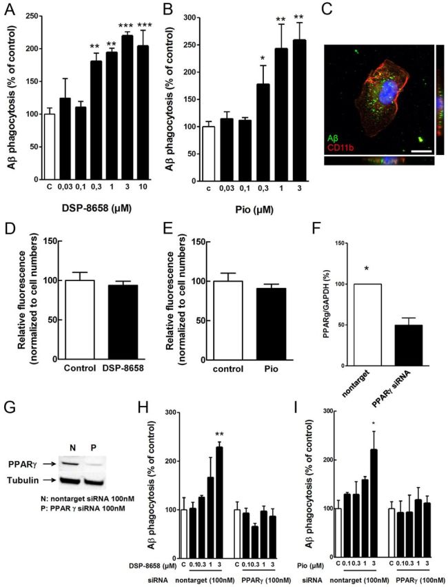
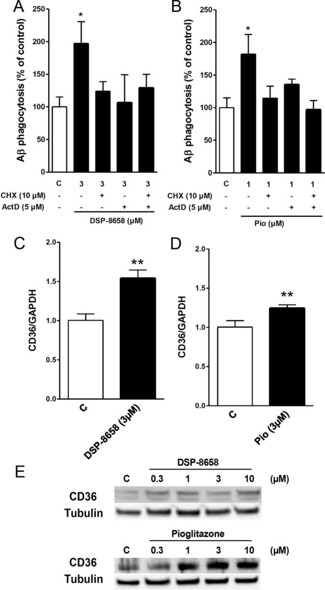
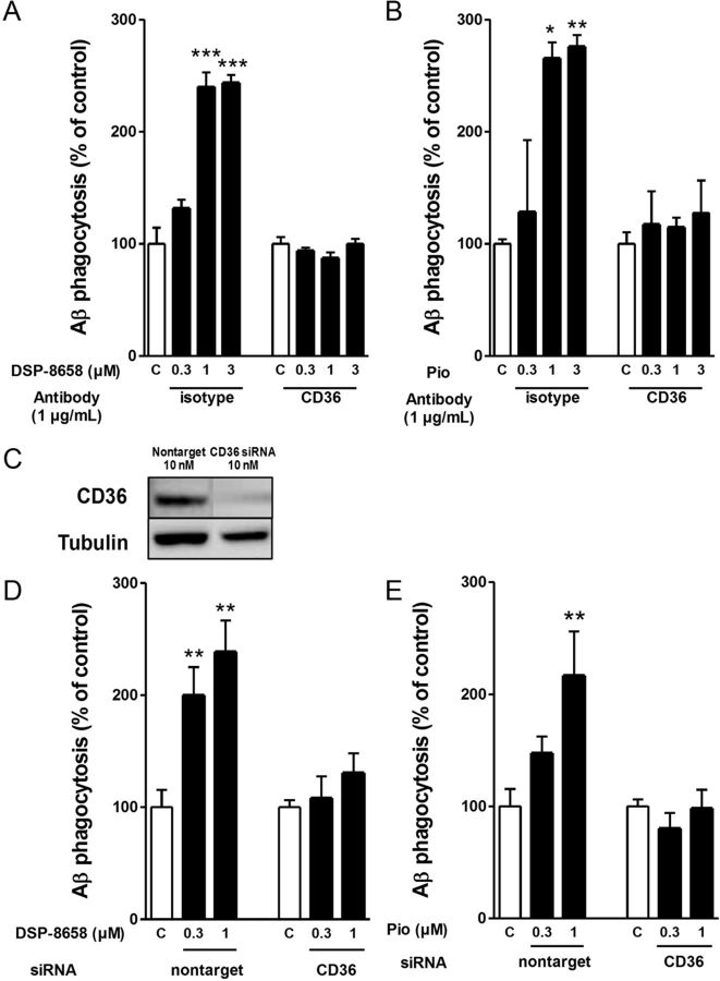
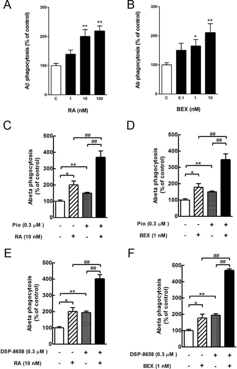
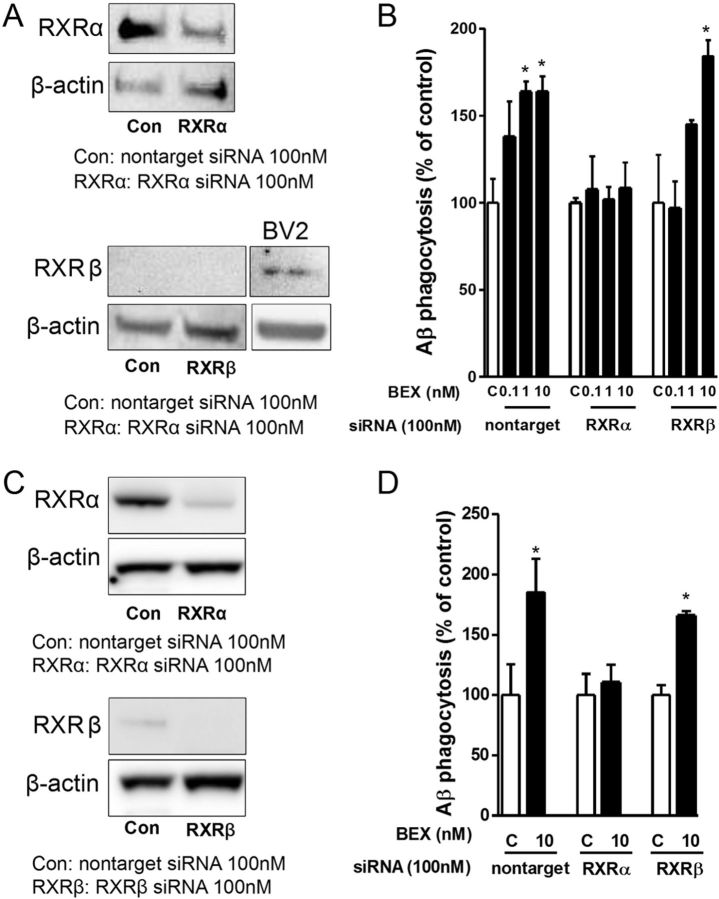
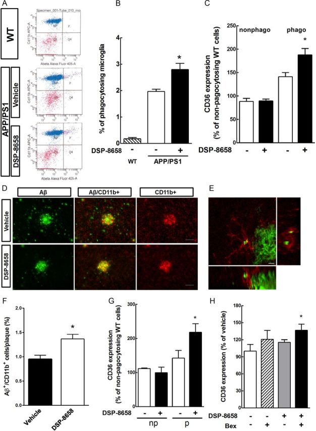
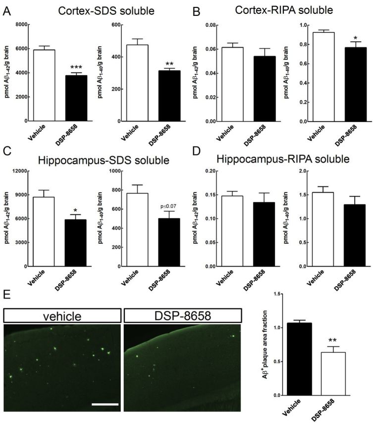
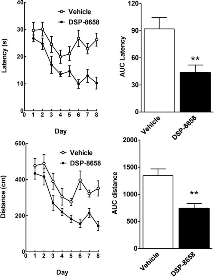
Alzheimer's disease (AD) is characterized by the extracellular deposition of amyloid-β (Aβ), neurofibrillary tangle formation, and a microglial-driven inflammatory response. Chronic inflammatory activation compromises microglial clearance functions. Because peroxisome proliferator-activated receptor γ (PPARγ) agonists suppress inflammatory gene expression, we tested whether activation of PPARγ would also result in improved microglial Aβ phagocytosis. The PPARγ agonist pioglitazone and a novel selective PPARα/γ modulator, DSP-8658, currently in clinical development for the treatment of type 2 diabetes, enhanced the microglial uptake of Aβ in a PPARγ-dependent manner. This PPARγ-stimulated increase of Aβ phagocytosis was mediated by the upregulation of scavenger receptor CD36 expression. In addition, combined treatment with agonists for the heterodimeric binding partners of PPARγ, the retinoid X receptors (RXRs), showed additive enhancement of the Aβ uptake that was mediated by RXRα activation. Evaluation of DSP-8658 in the amyloid precursor protein/presenilin 1 mouse model confirmed an increased microglial Aβ phagocytosis in vivo, which subsequently resulted in a reduction of cortical and hippocampal Aβ levels. Furthermore, DSP-8658-treated mice showed improved spatial memory performance. Therefore, stimulation of microglial clearance by simultaneous activation of the PPARγ/RXRα heterodimer may prove beneficial in prevention of AD.


Figures

Figure 1.
Figure 1.
Effect of PPARγ agonist on Aβ phagocytosis in primary microglia. Rat primary microglia were incubated with increasing concentrations of DSP-8658 or pioglitazone in the presence of FAM-Aβ (0.5 μm). After 4 h exposure to Aβ, intracellular levels of Aβ were determined. Both DSP-8658 (A) and pioglitazone (B) enhanced the uptake of Aβ in rat primary microglia in a concentration-dependent manner (mean ± SEM of n = 3, *p < 0.05, **p < 0.01, ***p < 0.001, one-way ANOVA, Tukey's post hoc test). C, Intracellular uptake of Aβ1–42 was confirmed by immunocytochemical staining of Aβ using antibody IC16 and the cell-surface antigen CD11b using antibody MCA711. Scale bar, 10 μm. Rat primary microglia were incubated for 30 min with FAM-Aβ in the presence of 0.3 μm DSP-8658 (D) or 0.3 μm pioglitazone (pio) (E) (mean ± SEM of n = 3). PPARγ mRNA (F) and protein levels (G) in primary microglia transfected with siRNA for PPARγ (mean ± SEM of n = 3, *p < 0.05, Student's t test). Primary microglia transfected with siRNA for PPARγ or nontarget were incubated with DSP-8658 (H) or pioglitazone (I) in the presence of FAM-Aβ (0.5 μm). DSP-8658 and pioglitazone enhanced Aβ uptake at 3 μm in nontarget control siRNA-treated primary microglia, although the same treatments failed to increase Aβ phagocytosis in PPARγ siRNA-treated primary microglia (mean ± SEM of n = 3, *p < 0.05, **p < 0.01, one-way ANOVA, Tukey's post hoc test).
Figure 2.
Figure 2.
Effects of PPARγ agonist on CD36 expression in primary microglia. Primary microglia was coincubated with inhibitors of transcription [actinomycin D (ActD), 5 μm] or translation [cycloheximide (CHX), 10 μm] in the presence of FAM-Aβ (0.5 μm). After 4 h, intracellular levels of Aβ were determined. Aβ phagocytosis by DSP-8658 (A) or pioglitazone (B) was abolished by coincubation with the respective inhibitors. The data show the intracellular Aβ uptake as percentage of control (mean ± SEM of n = 3, *p < 0.05, one-way ANOVA, Tukey's post hoc test). C–E, Primary microglia was incubated with DSP-8658 or pioglitazone for 6 h, and CD36 mRNA and protein levels were detected. CD36 mRNA was upregulated in response to DSP-8658 (C) or pioglitazone (D), followed by an increase of the respective protein levels in a concentration-dependent manner (E) (mean ± SEM of n = 3, **p < 0.01, Student's t test).
Figure 3.
Figure 3.
CD36 mediates the PPARγ-stimulated increase in microglial Aβ uptake. Primary microglia was incubated for 4 h with either DSP-8658 (A) or pioglitazone (B; Pio) in the presence of CD36 antibody or isotype control. After 4 h, intracellular Aβ was determined. PPARγ agonist-stimulated Aβ phagocytosis was suppressed by coincubation with CD36 antibody (mean ± SEM of n = 3, *p < 0.05, **p < 0.01, ***p < 0.001, one-way ANOVA, Tukey's post hoc test). C, Protein levels of CD36 in primary microglia transfected with siRNA for CD36. D, E, Primary microglia transfected with siRNA for CD36 or nontarget were incubated with DSP-8658 or pioglitazone in the presence of FAM-labeled Aβ1–42 (0.5 μm). After 4 h, intracellular level of Aβ was determined. Transfection of cell with CD36 siRNA blocked the PPARγ agonist-stimulated uptake of Aβ. The data show the intracellular Aβ uptake as percentage of control (mean ± SEM of n = 3, **p < 0.01, one-way ANOVA, Tukey's post hoc test).
Figure 4.
Figure 4.
Additive effects dual activation of PPARγ and RXR in primary microglia. Primary microglia were incubated with indicated concentrations of retinoic acid (A; RA) or bexarotene (B; BEX) in the presence of FAM-Aβ (0.5 μm). After 4 h expose to Aβ, intracellular levels of Aβ were determined. RXR agonists enhanced the uptake of Aβ in rat primary microglia in a concentration-dependent manner. The data show the intracellular Aβ uptake as percentage of control (mean ± SEM of n = 3, *p < 0.05, **p < 0.01, one-way ANOVA, Dunnett's multiple comparison test). C–F, Primary microglia was incubated with the combination of pioglitazone (0.3 μm) (C, D; Pio) or DSP-8658 (0.3 μm) (E, F) with the RXR agonists retinoic acid (10 nm) and bexarotene (1 nm) in the presence of FAM-Aβ (0.5 μm). After 4 h expose to Aβ, intracellular levels of Aβ were determined. Coincubation of PPARγ agonists with RXR agonists showed an additive enhancement on Aβ uptake. The data show the intracellular Aβ uptake as percentage of control (means ± SEM of n = 3, *p < 0.05, **p < 0.01 vs control, ##p < 0.01 vs DSP-8658, pioglitazone, retinoic acid, or bexarotene alone, one-way ANOVA, Tukey's post hoc test).
Figure 5.
Figure 5.
Effects of RXRα or RXRβ suppression on Aβ phagocytosis in rat primary microglia. A, RXRα and RXRβ detection in rat primary microglia transfected with siRNA for RXRα, RXRβ, or nontarget. B, Primary microglia transfected with siRNA for RXRα or RXRβ were incubated with bexarotene (BEX) in the presence of FAM-Aβ (0.5 μm). After 4 h expose to Aβ, intracellular level of Aβ was determined. Phagocytosis was only suppressed by RXRα siRNA. The data show the intracellular Aβ uptake as percentage of control (mean ± SEM of n = 3, *p < 0.05 vs control, one-way ANOVA, Tukey's post hoc test). C, RXRα and RXRβ detection in murine primary microglia transfected with siRNA for RXRα, RXRβ, or nontarget. D, Primary murine microglia transfected with siRNA for RXRα or RXRβ were incubated with bexarotene (BEX) in the presence of FAM-Aβ (0.5 μm). After 4 h exposure to Aβ, the intracellular level of Aβ was determined. Data show the intracellular Aβ uptake as percentage of control (mean ± SEM of n = 3, *p < 0.05 vs control, one-way ANOVA, Tukey's post hoc test).
Figure 6.
Figure 6.
DSP-8658 induces in vivo Aβ phagocytosis and recruitment of microglia to Aβ plaques. APP/PS1 transgenic mice received either DSP-8658 or vehicle containing chow for 3 months. A, Scatter blots of CD11b+/CD45+ isolated microglia after peripheral application of methoxy-X04. Background signal was determined using wild-type (WT) control mice. General microglial background was determined by analysis of unstained, non-injected wild-type mice. B, The percentage of methoxy-X04+/CD11b+/CD45+ cells to CD11b+/CD45+ cells was calculated (mean ± SEM of n = 4, *p < 0.05, Student's t test). C, FACS analysis for CD36 expression was performed in non-phagocytosing (nonphago) and phagocytosing (phago) cells (mean ± SEM of n = 4, *p < 0.05, Student's t test). D, Confocal laser-scanning microscopy detected a higher number of Aβ-plaque-associated and Aβ-containing microglial cells in DSP-8658-treated APP/PS1 transgenic mice compared with vehicle-treated APP/PS1 controls. Scale bar, 50 μm. E, Colocalization of Aβ/Cd11b within microglia visualized by confocal laser-scanning microscopy. Scale bar, 5 μm. F, Ten randomly chosen plaque areas were evaluated for Aβ/Cd11b colocalization per animal (mean ± SEM of n = 4, *p < 0.05, Student's t test). G, APP/PS1 transgenic mice were treated with DSP-8658 for 3 d and injected with methoxy-X04 3 h before analysis. After isolation of microglia, CD36 expression was determined by flow cytometry in non-phagocytosing (np) and phagocytosing (p) cells. Values were normalized to the CD36 expression in untreated microglia from age-matched wild-type mice (mean ± SEM of n = 4, *p < 0.05, Student's t test). H, APP/PS1 transgenic mice were treated with DSP-8658 or bexarotene (Bex) and the combination of these compounds for 3 d. Isolated phagocytosing microglia were analyzed by FACS for CD36 expression per animal (mean ± SEM of n = 4, *p < 0.05, Student's t test).
Figure 7.
Figure 7.
DSP-8658 reduces Aβ burden in the APP/PS1 transgenic mice. APP/PS1 transgenic mice were treated with DSP-8658 in the diet for 3 months. After treatment, cortex and hippocampus were excised and protein was extracted. Concentrations of Aβ1–40 and Aβ1–42 in SDS-soluble (A, C) and RIPA-soluble (B, D) fractions from cortex and hippocampus of APP/PS1 mice were determined by sandwich ELISA. DSP-8658 reduced SDS-soluble Aβ1–40 and Aβ1–42 in cortex and SDS-soluble Aβ1–42 in hippocampus compared with control (mean ± SEM of n = 4, *p < 0.05, **p < 0.01, Student's t test). E, Aβ deposition in the cortex was evaluated by thioflavin S staining and calculated as Aβ-positive area fraction (mean ± SEM of n = 4, *p < 0.05, Student's t test). Scale bar, 250 μm.
Figure 8.
Figure 8.
DSP-8658 treatment improved spatial memory learning of APP/PS1 transgenic mice. APP/PS1 transgenic mice were treated with DSP-8658 for 3 months. After treatment, all animals were subjected to the Morris water maze test. The time needed to reach the hidden platform (latency, seconds) and distance traveled (distance, centimeters) are depicted over 8 consecutive days. Integrated time and distance traveled (AUC) were determined for the whole observation period. Data represent means ± SEM (n = 9 for vehicle and n = 10 for DSP-8658, **p < 0.01, Student's t test).

Comment in

References

    1. Bamberger ME, Harris ME, McDonald DR, Husemann J, Landreth GE. A cell surface receptor complex for fibrillar β-amyloid mediates microglial activation. J Neurosci. 2003;23:2665–2674. - PMC - PubMed
    1. Bolmont T, Haiss F, Eicke D, Radde R, Mathis CA, Klunk WE, Kohsaka S, Jucker M, Calhoun ME. Dynamics of the microglial/amyloid interaction indicate a role in plaque maintenance. J Neurosci. 2008;28:4283–4292. - PMC - PubMed
    1. Colton CA, Mott RT, Sharpe H, Xu Q, Van Nostrand WE, Vitek MP. Expression profiles for macrophage alternative activation genes in AD and in mouse models of AD. J Neuroinflammation. 2006;3:27. - PMC - PubMed
    1. Combs CK, Johnson DE, Karlo JC, Cannady SB, Landreth GE. Inflammatory mechanisms in Alzheimer's disease: inhibition of beta-amyloid-stimulated proinflammatory responses and neurotoxicity by PPARγ agonists. J Neurosci. 2000;20:558–567. - PMC - PubMed
    1. Coraci IS, Husemann J, Berman JW, Hulette C, Dufour JH, Campanella GK, Luster AD, Silverstein SC, El-Khoury JB. CD36, a class B scavenger receptor, is expressed on microglia in Alzheimer's disease brains and can mediate production of reactive oxygen species in response to beta-amyloid fibrils. Am J Pathol. 2002;160:101–112. - PMC - PubMed

Publication types

MeSH terms