

Characteristic distributions of intracerebral hemorrhage-associated diffusion-weighted lesions
- PMID: 23197745
- PMCID: PMC3578378
- DOI: 10.1212/WNL.0b013e318278b66f
Characteristic distributions of intracerebral hemorrhage-associated diffusion-weighted lesions
Erratum in
- Neurology. 2013 Oct;81(15):1367
Abstract
Objectives: To determine whether small diffusion-weighted imaging (DWI) lesions occur beyond the acute posthemorrhage time window in patients with intracerebral hemorrhage (ICH) and to characterize their spatial distribution in patients with lobar and deep cerebral hemorrhages.
Methods: In this cross-sectional study, we retrospectively analyzed 458 MRI scans obtained in the acute (≤ 7 days after ICH) or nonacute (>14 days after ICH) phases from 392 subjects with strictly lobar (n = 276) and deep (n = 116) ICH (48.7% women; mean age 72.8 ± 11.7 years). DWI, apparent diffusion coefficient maps, fluid-attenuated inversion recovery, and T2* MRIs were reviewed for the presence and location of DWI lesions.
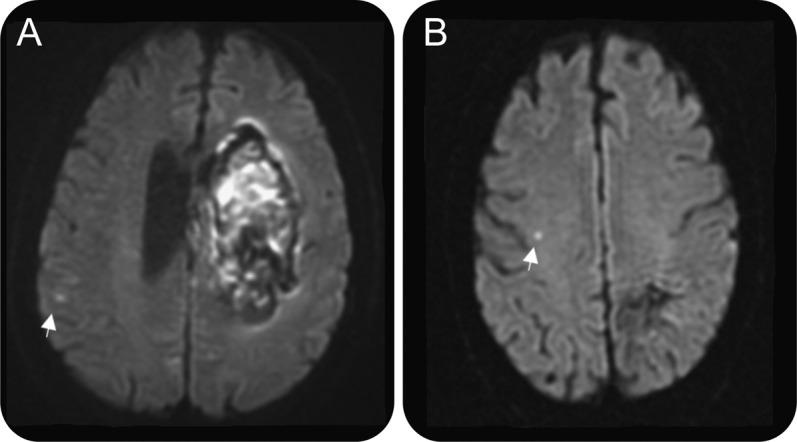
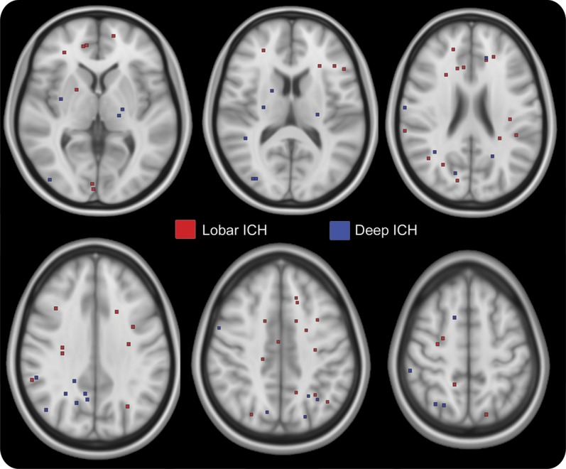
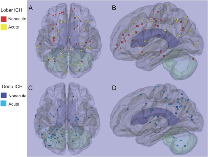
Results: We identified 103 DWI hyperintense lesions on scans from 62 subjects, located mostly in lobar brain regions (90 of 103, 87.4%). The lesions were not uniformly distributed throughout the brain lobes; patients with strictly lobar ICH had relative overrepresentation of lesions in frontal lobe, and patients with deep ICH in parietal lobe (p = 0.002). Although the frequency of DWI lesions tended to be greater on scans performed within 7 days after ICH (39 of 214, 18.2%), they continued at high frequency in the nonacute period as well (23 of 178, 12.9%, odds ratio 1.5, 95% confidence interval 0.86-2.6 for acute vs nonacute). There was also no difference in frequency of lesions on acute and nonacute scans among 66 subjects with MRIs in both time periods (8 of 66 acute, 10 of 66 nonacute, odds ratio 0.77, 95% confidence interval 0.25-2.4).
Conclusions: The high frequency of DWI lesions beyond the acute post-ICH period and their characteristic distributions suggest that they are products of the small vessel diseases that underlie ICH.
Figures
References
-
- Prabhakaran S, Gupta R, Ouyang B, et al. Acute brain infarcts after spontaneous intracerebral hemorrhage: a diffusion-weighted imaging study. Stroke 2010;41:89–94 - PubMed
-
- Gregoire SM, Charidimou A, Gadapa N, et al. Acute ischaemic brain lesions in intracerebral haemorrhage: multicentre cross-sectional magnetic resonance imaging study. Brain 2011;134:2376–2386 - PubMed
Publication types
MeSH terms
Grants and funding
LinkOut - more resources
Full Text Sources
Other Literature Sources