. 2012 Apr;1(4):309-21.
doi: 10.5966/sctm.2011-0010. Epub 2012 Apr 2.

Sp1 transcription factor interaction with accumulated prelamin a impairs adipose lineage differentiation in human mesenchymal stem cells: essential role of sp1 in the integrity of lipid vesicles

Affiliations

Sp1 transcription factor interaction with accumulated prelamin a impairs adipose lineage differentiation in human mesenchymal stem cells: essential role of sp1 in the integrity of lipid vesicles

Garbiñe Ruiz de Eguino et al. Stem Cells Transl Med. 2012 Apr.

Abstract

Lamin A (LMNA)-linked lipodystrophies may be either genetic (associated with LMNA mutations) or acquired (associated with the use of human immunodeficiency virus protease inhibitors [PIs]), and in both cases they share clinical features such as anomalous distribution of body fat or generalized loss of adipose tissue, metabolic alterations, and early cardiovascular complications. Both LMNA-linked lipodystrophies are characterized by the accumulation of the lamin A precursor prelamin A. The pathological mechanism by which prelamin A accumulation induces the lipodystrophy associated phenotypes remains unclear. Since the affected tissues in these disorders are of mesenchymal origin, we have generated an LMNA-linked experimental model using human mesenchymal stem cells treated with a PI, which recapitulates the phenotypes observed in patient biopsies. This model has been demonstrated to be a useful tool to unravel the pathological mechanism of the LMNA-linked lipodystrophies, providing an ideal system to identify potential targets to generate new therapies for drug discovery screening. We report for the first time that impaired adipogenesis is a consequence of the interaction between accumulated prelamin A and Sp1 transcription factor, sequestration of which results in altered extracellular matrix gene expression. In fact, our study shows a novel, essential, and finely tuned role for Sp1 in adipose lineage differentiation in human mesenchymal stem cells. These findings define a new physiological experimental model to elucidate the pathological mechanisms LMNA-linked lipodystrophies, creating new opportunities for research and treatment not only of LMNA-linked lipodystrophies but also of other adipogenesis-associated metabolic diseases.
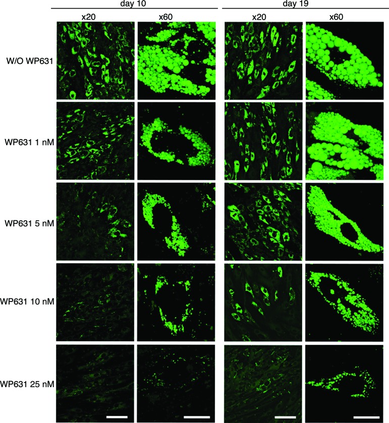

Figures

Figure 1.
Figure 1.
Prelamin A accumulation and altered chromatin organization in TPV-treated human mesenchymal stem cells (hMSCs). (A): Western blot of prelamin A. Farnesyl transferase inhibitor treatment is shown as positive control. Lamin C was used as a loading control. (B): Prelamin A staining by immunofluorescence. Scale bar = 50 μm. (C, D): Electron micrographs showing heterochromatin aggregates (small arrows) and lipid bilayer (large arrows) carried out in three different BM-hMSCs. Aberrant condensation of heterochromatin is shown in the lower panels in (D). Scale bars = 2 nm (C) and 500 nm (D). Abbreviations: BM, bone marrow; cont, control; DAPI, 4′,6-diamidino-2-phenylindole; TPV, tipranavir.
Figure 2.
Figure 2.
TPV-treated human mesenchymal stem cells (hMSCs) show reduced proliferation, premature senescence, and repression of the retinoic acid pathway. (A): Population doubling levels carried out in duplicate in four different BM-hMSCs. (B): Annexin V-FITC Propidium Iodide staining. (C): Senescence-associated β-galactosidase activity. Senescent cells were counted in randomly selected fields and are expressed as a percentage of total cells. Scale bars = 500 μm. (D): RARE and NF-κB luciferase reporter activity. (B–D): Studies were performed in triplicate in two different BM-hMSCs. RARE reporter analyses were carried out in duplicate in four different BM-hMSCs. Abbreviations: BM, bone marrow; NF, nuclear factor; RARE, retinoic acid response element; TPV, tipranavir.
Figure 3.
Figure 3.
Prelamin A accumulation induced by TPV impairs human mesenchymal stem cell (hMSC) differentiation into adipogenic lineage. (A, C): Immunofluorescence staining for prelamin A (red) and lipid droplets (green) in hMSC-derived adipocytes. Scale bars = 100 μm (A) and 20 μm (C). (B): Oil Red O staining in hMSC-derived adipocytes in late adipogenesis. (D): DAPI-stained immunofluorescence images showing nuclear blebbing carried out in three different bone marrow hMSC-derived adipocytes. Abbreviations: BM, bone marrow; DAPI, 4′,6-diamidino-2-phenylindole; TPV, tipranavir.
Figure 4.
Figure 4.
Prelamin A colocalizes with Sp1 in late adipogenesis, affecting Sp1 transcription factor activity. (A): Proximity ligation assay for prelamin A and Sp1. Red dots correspond to a protein-protein physical interaction. Scale bar = 20 μm. (B): Sp1 reporter vector assay performed in triplicate in bone marrow-derived human mesenchymal stem cells (hMSCs) and in duplicate in different bone marrow-derived hMSCs. hMSCs were nucleofected; 24 hours later, adipogenesis was induced for 72 hours, and then firefly and Renilla activities were measured. Abbreviations: BM, bone marrow; TPV, tipranavir.
Figure 5.
Figure 5.
Sp1 activity is essential for lipid vesicle integrity in human mesenchymal stem cells (hMSCs). hMSCs were differentiated to adipocytes, and 10 or 19 days after induction, WP631 was added to the medium at different concentrations for 24 hours. Immunofluorescence was performed with BODIPY staining (at day 12 or 21). Scale bars = 100 μm (×20 columns) and 20 μm (×60 columns). Abbreviation: W/O, without.

Similar articles

Cited by

References

    1. Mounkes LC, Stewart CL. Aging and nuclear organization: Lamins and progeria. Curr Opin Cell Biol. 2004;16:322–327. - PubMed
    1. Burke B, Stewart CL. The laminopathies: The functional architecture of the nucleus and its contribution to disease. Annu Rev Genomics Hum Genet. 2006;7:369–405. - PubMed
    1. Hegele RA. Monogenic forms of insulin resistance: Apertures that expose the common metabolic syndrome. Trends Endocrinol Metab. 2003;14:371–377. - PubMed
    1. Mattout A, Dechat T, Adam SA, et al. Nuclear lamins, diseases and aging. Curr Opin Cell Biol. 2006;18:335–341. - PubMed
    1. Shackleton S, Lloyd DJ, Jackson SN, et al. LMNA, encoding lamin A/C, is mutated in partial lipodystrophy. Nat Genet. 2000;24:153–156. - PubMed

Publication types

MeSH terms