Comparative Study
. 2012 May;1(5):396-402.
doi: 10.5966/sctm.2011-0061. Epub 2012 May 8.

Amyotrophic lateral sclerosis model derived from human embryonic stem cells overexpressing mutant superoxide dismutase 1

Affiliations
Comparative Study

Amyotrophic lateral sclerosis model derived from human embryonic stem cells overexpressing mutant superoxide dismutase 1

Tamaki Wada et al. Stem Cells Transl Med. 2012 May.

Abstract

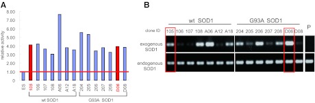
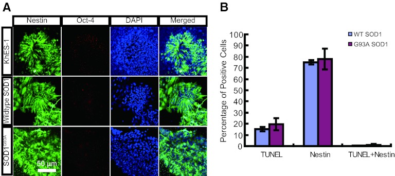
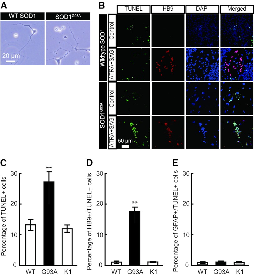
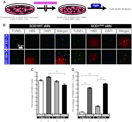
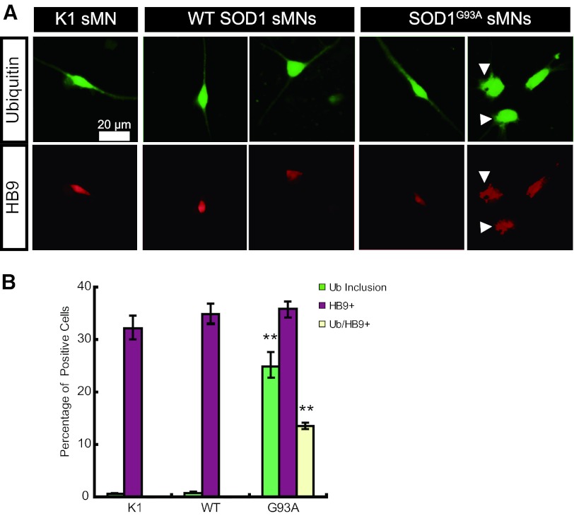
The generation of amyotrophic lateral sclerosis (ALS) disease models is an important subject for investigating disease mechanisms and pharmaceutical applications. In transgenic mice, expression of a mutant form of superoxide dismutase 1 (SOD1) can lead to the development of ALS that closely mimics the familial type of ALS (FALS). Although SOD1 mutant mice show phenotypes similar to FALS, dissimilar drug responses and size differences limit their usefulness to study the disease mechanism(s) and identify potential therapeutic compounds. Development of an in vitro model system for ALS is expected to help in obtaining novel insights into disease mechanisms and discovery of therapeutics. We report the establishment of an in vitro FALS model from human embryonic stem cells overexpressing either a wild-type (WT) or a mutant SOD1 (G93A) gene and the evaluation of the phenotypes and survival of the spinal motor neurons (sMNs), which are the neurons affected in ALS patients. The in vitro FALS model that we developed mimics the in vivo human ALS disease in terms of the following: (a) selective degeneration of sMNs expressing the G93A SOD1 but not those expressing the WT gene; (b) susceptibility of G93A SOD1-derived sMNs to form ubiquitinated inclusions; (c) astrocyte-derived factor(s) in the selective degeneration of G93A SOD1 sMNs; and (d) cell-autonomous, as well as non-cell-autonomous, dependent sMN degeneration. Thus, this model is expected to help unravel the disease mechanisms involved in the development of FALS and also lead to potential drug discoveries based on the prevention of neurodegeneration.


Figures

Figure 1.
Figure 1.
Establishment of SOD1-overexpressing human embryonic stem cell lines. (A): Relative SOD activity in undifferentiated human embryonic stem cells (hESCs) ectopically expressing wild-type SOD1 or G93A SOD1. (B): Reverse transcription-polymerase chain reaction analysis of exogenous and endogenous SOD1 expression in hESC-derived neurons. Red bars (A) and open rectangles (B) show clones selected for further analysis. Abbreviations: ID, identification number; P, parent hESC line KhES-1; SOD, superoxide dismutase; wt, wild-type.
Figure 2.
Figure 2.
Phenotypic analysis of SOD1-overexpressing human ESC-derived neural progenitor cells. (A): Immunocytochemical staining by Nestin (neural marker; green) and Oct-4 (ESC marker; red) of KhES-1, WT SOD1, and G93A SOD1 cells at the end of the neural progenitor formation stage. (B): Quantitation of TUNEL-positive, Nestin-positive, and double-positive cell ratios. Abbreviations: DAPI, 4′,6-diamidino-2-phenylindole; SOD, superoxide dismutase; TUNEL, terminal deoxynucleotidyl transferase dUTP nick-end labeling; WT, wild-type.
Figure 3.
Figure 3.
Phenotypic analysis of SOD1-overexpressing human ESC (hESC)-derived spinal motor neurons. (A): Morphologically unhealthy cells were observed in spinal motor neurons (sMNs) derived from G93A SOD1-overexpressing hESCs, whereas WT SOD1 hESC-derived sMNs showed normal morphology. (B): Immunocytochemical staining of cells treated with or without sMN inducers (ATRA and SAG). Both TUNEL (green) and HB9 (red)-positive sMNs were observed in ATRA/SAG-treated G93A SOD1-overexpressing hESCs. (C): Total TUNEL-positive cell ratio. (D): HB9/TUNEL double-positive cell ratio. (E): GFAP/TUNEL double-positive cell ratio. These data indicated sMN-specific cell death in G93A SOD1 hESC-derived sMNs. Approximately 900 cells were used for calculating the ratio of immunopositive cells (TUNEL, HB9, and GFAP). Mean ± SEM; n = 9; Steel-Dwass test; ∗∗, p < .01. Abbreviations: ATRA, all-trans retinoic acid; DAPI, 4′,6-diamidino-2-phenylindole; GFAP, glial fibrillary acidic protein; K1, KhES-1; SOD, superoxide dismutase; TUNEL, terminal deoxynucleotidyl transferase dUTP nick-end labeling; WT, wild-type.
Figure 4.
Figure 4.
G93A SOD1-overexpressing human ESC (hESC)-derived astrocyte supernatant-induced sMN death. (A): Experimental procedures to analyze the effect of astrocyte-CM on sMN cell death. (B): Immunocytochemical staining of astrocyte-CM-treated sMNs from either WT SOD1 or G93A SOD1-overexpressing hESCs. (C, D): Quantitative analysis of TUNEL-positive dead cells (C) and HB9/TUNEL-double-positive dead sMNs (D). Mean ± SEM; n = 9; Steel-Dwass test; ∗∗, p < .01. Abbreviations: AS CM, astrocyte-conditioned medium; CM, conditioned medium; DAPI, 4′,6-diamidino-2-phenylindole; DIV, days in vitro; sMN, spinal motor neuron; SOD, superoxide dismutase; TUNEL, terminal deoxynucleotidyl transferase dUTP nick-end labeling; WT, wild-type.
Figure 5.
Figure 5.
Ubiquitin staining of SOD1 human ESC-derived sMNs. (A): Immunocytochemical staining of ubiquitin (green) and HB9 (red). An abnormal staining pattern was observed in G93A SOD1-expressing sMNs. Arrowheads show rough staining of ubiquitin or HB9, which might be abnormal ubiquitin inclusions. (B): Quantitative analysis of cells with ubiquitin inclusions (green bars), HB9-positive sMNs (purple bars), and sMNs with ubiquitin inclusions (yellow bars). Mean ± SEM; n = 9; Steel-Dwass test; ∗∗, p < .01. Abbreviations: sMN, spinal motor neuron; SOD, superoxide dismutase; Ub, ubiquitin; WT, wild-type.

Similar articles

Cited by

References

    1. Ozdinler PH, Benn S, Yamamoto TH, et al. Corticospinal motor neurons and related subcerebral projection neurons undergo early and specific neurodegeneration in hSOD1G93A transgenic ALS mice. J Neurosci. 2011;31:4166–4177. - PMC - PubMed
    1. Kiernan MC, Vucic S, Cheah BC, et al. Amyotrophic lateral sclerosis. Lancet. 2011;377:942–955. - PubMed
    1. Kwiatkowski TJ, Jr, Bosco DA, Leclerc AL, et al. Mutations in the FUS/TLS gene on chromosome 16 cause familial amyotrophic lateral sclerosis. Science. 2009;323:1205–1208. - PubMed
    1. Vance C, Rogelj B, Hortobágyi T, et al. Mutations in FUS, an RNA processing protein, cause familial amyotrophic lateral sclerosis type 6. Science. 2009;323:1208–1211. - PMC - PubMed
    1. Wegorzewska I, Bell S, Cairns NJ, et al. TDP-43 mutant transgenic mice develop features of ALS and frontotemporal lobar degeneration. Proc Natl Acad Sci USA. 2009;106:18809–18814. - PMC - PubMed

Publication types