. 2012 Dec 26;109(52):E3678-86.
doi: 10.1073/pnas.1214572109. Epub 2012 Nov 29.

Insulator-like pairing elements regulate silencing and mutually exclusive expression in the malaria parasite Plasmodium falciparum

Affiliations

Insulator-like pairing elements regulate silencing and mutually exclusive expression in the malaria parasite Plasmodium falciparum

Inbar Avraham et al. Proc Natl Acad Sci U S A. .

Abstract

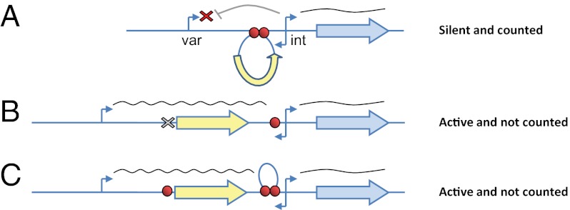
Plasmodium falciparum causes the deadliest form of human malaria. Its virulence is attributed to its ability to modify the infected RBC and to evade human immune attack through antigenic variation. Antigenic variation is achieved through tight regulation of antigenic switches between variable surface antigens named "P. falciparum erythrocyte membrane protein-1" encoded by the var multicopy gene family. Individual parasites express only a single var gene at a time, maintaining the remaining var genes in a transcriptionally silent state. Strict pairing between var gene promoters and a second promoter within an intron found in each var gene is required for silencing and counting of var genes by the mechanism that controls mutually exclusive expression. We have identified and characterized insulator-like DNA elements that are required for pairing var promoters and introns and thus are essential for regulating silencing and mutually exclusive expression. These elements, found in the regulatory regions of each var gene, are bound by distinct nuclear protein complexes. Any alteration in the specific, paired structure of these elements by either deletion or insertion of additional elements results in an unregulated var gene. We propose a model by which silencing and mutually exclusive expression of var genes is regulated by the precise arrangement of insulator-like DNA pairing elements.


Conflict of interest statement

The authors declare no conflict of interest.

Figures

Fig. 1.
Fig. 1.
A DNA element found in the PbDT 3′ UTR disrupts the intron’s ability to silence a var promoter. Stably transfected parasite lines carried constructs mimicking var gene structure with luciferase as a reporter gene for var promoter expression and hdhfr as a positive selectable marker expressed by the var intron. (A) (Upper) Schematic of the pVLhIDb episome that contains a var promoter paired with a var intron. This var promoter is properly regulated and silent by default. (Lower) Schematic of the pVLbIDh episome, which is identical to pVLhIDb except for the replacement of the 3′ UTR that terminates luciferase. This replacement resulted in activation of the var promoter, which no longer is recognized by the mechanism that controls mutually exclusive expression. (B) Results of pVLhIDb (Upper) and pVLbIDh (Lower) in the DC-J parasite line. Quantification of the ratio between var promoter and intron by qRT-PCR (Left). Steady-state mRNA levels of each individual var gene measured by qRT-PCR are presented as copy number relative to the housekeeping genes arginyl-tRNA synthetase (PFL0900c) (Center) and the levels of luciferase expression (Right). The D10 parasite line (22) constitutively expressing luciferase from an endogenous var promoter was used as a positive control. All values presented are the average of at least two biological replicates. Error bars represent SE. (C) A similar TG-rich DNA element is found within the PbDT 3′ UTR, var promoter, and var intron. In the schematic of the construct, the TG-rich element is shown as a red circle, the PbDT 3′ UTR is boxed, and the hrp2 3′ UTR is shown as a bold black line.
Fig. 2.
Fig. 2.
Bioinformatic analysis of the entire var gene family revealed a unique TG-rich DNA motif in all var introns and 5′UTRs. The MEME motif-finding algorithm was used for the initial screen for TG-rich DNA motifs on all var introns of the 3D7 parasite line. Three T[G/A] motifs were identified, which were termed “M1-3” and are marked with light blue, blue, and red, respectively. The MAST algorithm was used to locate their presence in the 5′ UTRs of all var genes. This screen was limited to sequences with at least 25% G/C content. The presentation of these motifs above or below the genes indicates their orientation on the DNA strands.
Fig. 3.
Fig. 3.
DNA PEs regulate silencing and mutually exclusive expression of var genes through specific promoter–promoter interaction. The deletion of a PE or insertion of an extra PE results in the activation of a var gene. In the schematic of the construct, the TG-rich element is shown as a red circle, the PbDT 3′ UTR is boxed, and the hrp2 3′ UTR is shown as a bold black line. (A) (Upper) Schematic of the pVΔ102LhIDb in which the last 102 bp of the var promoter containing the PE were deleted. (Lower) Schematic of the pVLhPEIDh episome, which is identical to pVLhIDb except that an additional PE is inserted upstream of the var intron. These modifications resulted in activation of the var promoter, which no longer is recognized by the mechanism that controls mutually exclusive expression. (B) Results of pVΔ102LhIDb (Upper) and pVLhPEIDb (Lower) in the DC-J parasite line. Quantification of the ratio between var promoter and intron by qRT-PCR (Left), steady-state mRNA levels of each individual var gene measured by qRT-PCR presented as relative copy number to the housekeeping genes arginyl-tRNA synthetase (PFL0900c) (Center), and the levels of luciferase expression (Right). The D10 parasite line (22) constitutively expressing luciferase from an endogenous var promoter was used as a positive control. (C) The interaction between the two DNA elements is specific to var genes. Schematic of the pHLhIDb (Upper) and pHPELhIDb (Lower) constructs made by replacing the var promoter from pVLhIDb with the hrp2 promoter with or without the last 102 bp of the var promoter that contains the PE. The promoters of both plasmids were constitutively active simultaneously with the endogenous var gene in the DC-J parasite line. (D) Results of pHLhIDb (Upper) and pHPELhIDb (Lower) in the DC-J parasite line. (Left) Quantification of the ratio between var promoter and intron by qRT-PCR. Steady-state mRNA levels of each individual var gene measured by qRT-PCR are presented as relative copy number to the housekeeping genes arginyl-tRNA synthetase (PFL0900c) (Center) and the levels of luciferase expression (Right). The D10 parasite line (22) constitutively expressing luciferase from an endogenous var promoter was used as a positive control. All values presented are the average of at least two biological replicates. Error bars represent SEs.
Fig. 4.
Fig. 4.
The var PEs form a specific DNA–protein complex. (A) EMSA of extracts using a radiolabeled (32P) DNA ligand containing the PE of the var 5′UTR (Ups1) shows specific DNA–protein complex formation when incubated with a nuclear extract. Specific competition assays were performed with increasing concentrations of unlabeled DNA ligands (2×, 10×, and 25× of the labeled ligand, respectively) containing the PEs found in a var 5′UTR (Ups1), in a var intron (Int1), and in the PbDT-3′UTR (PbDT-3′). The DNA sequence from the hrp2-3′ UTR was used as nonspecific competitor (hrp2-3′). Blank, no protein was added; CE, cytoplasmic extract; NE, nuclear extract. (B) Phosphorimaging quantification of the EMSA data presented in A. The percentage of complex formation measured is presented relative to the value measured in the absence of competitive ligand, which was considered to be 100%. (C) Competition EMSA of radiolabeled Ups1 ligand with various mutated nonlabeled Ups1 ligands. The various mutated ligands are named “mut1-3,” “mut3-5,” and so forth, relative to the replacement of the original PE sequence with cytosine bases. The PE sequence is marked in bold. Base-pair exchange is underlined in the PE and is marked in gray in the flanking regions. Blank represents the original sequence of the PE and its flanking regions. (D) Phosphorimaging quantification of the EMSA data presented in C was performed, and the relative DNA-binding percentage was calculated as in B. (E) Schematic description of the importance of each nucleotide position in the PE for formation of the DNA–protein complex. Nucleotides shown in larger type have a great influence on binding.
Fig. P1.
Fig. P1.
A model for var gene regulation through promoter–promoter interaction, which is mediated by insulator-like DNA elements. (A) In a properly regulated var gene, the PEs (red circles) bind to specific nuclear proteins that mediate the interaction between the var and the intron promoters, thus enabling the intron promoter to silence the var promoter. Any alteration in the specific paired structure of these elements by either deletion (B) or insertion (C) of additional elements results in an unregulated var gene, which cannot be silenced and is not “counted” as a var gene for mutually exclusive expression.

Similar articles

Cited by

References

    1. WHO 2010. World Malaria Report 2010. (World Health Organization, Geneva)
    1. Baruch DI, Gormely JA, Ma C, Howard RJ, Pasloske BL. Plasmodium falciparum erythrocyte membrane protein 1 is a parasitized erythrocyte receptor for adherence to CD36, thrombospondin, and intercellular adhesion molecule 1. Proc Natl Acad Sci USA. 1996;93(8):3497–3502. - PMC - PubMed
    1. Smith JD, et al. Switches in expression of Plasmodium falciparum var genes correlate with changes in antigenic and cytoadherent phenotypes of infected erythrocytes. Cell. 1995;82(1):101–110. - PMC - PubMed
    1. Scherf A, et al. Antigenic variation in malaria: In situ switching, relaxed and mutually exclusive transcription of var genes during intra-erythrocytic development in Plasmodium falciparum. EMBO J. 1998;17(18):5418–5426. - PMC - PubMed
    1. Chen Q, et al. Developmental selection of var gene expression in Plasmodium falciparum. Nature. 1998;394(6691):392–395. - PubMed

Publication types

MeSH terms