. 2012 Dec 26;109(52):21208-15.
doi: 10.1073/pnas.1218836109. Epub 2012 Nov 28.

Folding of large multidomain proteins by partial encapsulation in the chaperonin TRiC/CCT

Affiliations

Folding of large multidomain proteins by partial encapsulation in the chaperonin TRiC/CCT

Florian Rüßmann et al. Proc Natl Acad Sci U S A. .

Abstract

The eukaryotic chaperonin, TRiC/CCT (TRiC, TCP-1 ring complex; CCT, chaperonin containing TCP-1), uses a built-in lid to mediate protein folding in an enclosed central cavity. Recent structural data suggest an effective size limit for the TRiC folding chamber of ∼70 kDa, but numerous chaperonin substrates are substantially larger. Using artificial fusion constructs with actin, an obligate chaperonin substrate, we show that TRiC can mediate folding of large proteins by segmental or domain-wise encapsulation. Single or multiple protein domains up to ∼70 kDa are stably enclosed by stabilizing the ATP-hydrolysis transition state of TRiC. Additional domains, connected by flexible linkers that pass through the central opening of the folding chamber, are excluded and remain accessible to externally added protease. Experiments with the physiological TRiC substrate hSnu114, a 109-kDa multidomain protein, suggest that TRiC has the ability to recognize domain boundaries in partially folded intermediates. In the case of hSnu114, this allows the selective encapsulation of the C-terminal ∼45-kDa domain and segments thereof, presumably reflecting a stepwise folding mechanism. The capacity of the eukaryotic chaperonin to overcome the size limitation of the folding chamber may have facilitated the explosive expansion of the multidomain proteome in eukaryotes.


Conflict of interest statement

The authors declare no conflict of interest.

Figures

Fig. 1.
Fig. 1.
TRiC-mediated folding of model multidomain proteins. (A) Domain structure of actin-fusion constructs. A, actin; GA, GFP-actin; AG, actin-GFP; BGA, BFP-GFP-actin; BAG, BFP-actin-GFP; BG, BFP-GFP. (B) Solubility of in vitro translation products. Actin-fusion proteins were synthesized in RRL (90 min, 30 °C) in the presence of [35S]-Met. Total translation reactions (T) were separated by centrifugation into soluble (S) and pellet (P) fractions and analyzed by SDS/PAGE and fluorography. Sections of gels containing the full-length proteins are shown. (C) Retention of actin-fusion constructs on DNase I beads. Adjacent lanes show total translation reactions (10% of input material) and the fraction bound to DNase I. The fraction of DNase I-bound BG (∼1% of total) represents nonspecific binding. (D) SDS/PAGE and fluorography of translation reactions expressing the proteins indicated. Reactions were analyzed after incubation without or with PK (83 μg/mL) for 10 min on ice. The PK-resistant actin fragment of ∼35 kDa derived from folded actin and the BFP/GFP domain are indicated. The positions of molecular weight (MW) markers are indicated in kDa. (E) Quantification of actin folding yield. The fraction of DNase I bound full-length protein (gray) and the relative intensity of the protease-resistant actin fragment (red) were quantified by densitometry. Averages ± SD from at least three independent experiments are shown. The intensity of the actin fragment (residues 48–375 of actin) relative to full-length actin was calculated by taking the number of methionine residues in actin and GFP/BFP into account.
Fig. 2.
Fig. 2.
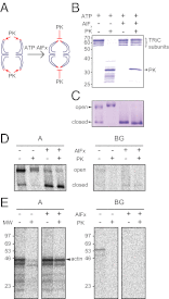
Stabilizing the closed state of TRiC. (A) Principle of protease protection assay. Binding of ATP·AlFx stabilizes TRiC in the closed state. The apical domains of the TRiC subunits, shown in red, are sensitive to proteolytic cleavage by PK in the open state, but not in the closed state. Note that protease cleavage in the apical domains does not affect complex assembly (33), as shown in C. (B) Protease protection of TRiC in the closed conformation. Purified bovine TRiC was incubated with or without ATP·AlFx (60 min at 30 °C), followed by treatment with PK as described in Materials and Methods. Reactions were analyzed by SDS/PAGE and Coomassie staining. (C) Analysis of TRiC by native PAGE and Coomassie staining. Samples were treated as in B. Open TRiC migrates more slowly than the closed form. The TRiC complex remains assembled after PK cleavage. (D) (Left) Native PAGE analysis of TRiC-bound [35S]-Met labeled actin. (Right) [35S]-Met labeled BFP-GFP fusion protein (BG) served as a control for the specificity of TRiC binding. Actin and BG were synthesized in RRL as in Fig. 1. Translation reactions were incubated with or without ATP·AlFx and subjected to PK treatment before native PAGE. Radiolabeled protein bound to open or closed TRiC was visualized by fluorography. (E) Reanalysis of TRiC-bound [35S]-Met actin and BG by SDS/PAGE. The regions of the native gel in D containing open and closed TRiC were excised and reanalyzed by SDS/PAGE and fluorography. The positions of molecular weight (MW) markers are indicated in kDa.
Fig. 3.
Fig. 3.
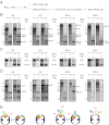
Encapsulation of actin fusion proteins by TRiC. (A) Flow diagram of the experiment. Translation reactions were treated with or without ATP·AlFx, followed by incubation with or without PK and analysis by SDS/PAGE or native PAGE and fluorography. TRiC-containing bands were excised from the native gel and reanalyzed by SDS/PAGE. (B) SDS/PAGE and fluorography of complete translation reactions of GA, AG, BGA, and BAG fusion proteins. The position and size of the full-length proteins as well as of protease-protected fragments are indicated. In the presence of ATP·AlFx, additional protease-protected bands are observed. The positions of molecular weight markers are indicated in kDa. (C) Native PAGE analysis of TRiC-bound actin fusion proteins. Translation reactions were treated as in B. The regions of native PAGE gels containing the TRiC complex are shown. (D) Reanalysis of TRiC-bound translation products by SDS/PAGE. Complexes of TRiC with actin fusion proteins were excised from native PAGE gels and separated by SDS/PAGE. The positions of molecular weight markers are indicated in kDa. (E) Proposed topology of TRiC-bound actin fusion proteins in the presence of ATP·AlFx. Red arrows show accessibility to protease. Note that in case of TRiC-bound BAG, the TRiC cavity fails to close completely.
Fig. 4.
Fig. 4.
Interaction of the large substrate protein hSnu114 with TRiC. (A) Structural model of hSnu114. (Upper) Predicted domain structure of hSnu114, based on the crystal structure of the homologous yeast protein, eEF2 (56). The domain nomenclature of eEF2 is used, and residue numbers corresponding to domain boundaries are indicated. (Lower) An hSnu114 structural model from the Swiss-Model Repository in ribbon representation (69). (B) Full-length hSnu114 [1–972] and fragments hSnu114 [1–580] and hSnu114 [581–972] were synthesized in RRL. Complete translation reactions were incubated with or without ATP·AlFx, followed by protease protection assay and analysis by SDS/PAGE and fluorography, as described in Fig. 3B. The position and size of the full-length translation products as well as of protease-protected fragments are indicated in kDa (K). The positions of molecular weight markers are indicated on the left as in Fig. 1D. (C) Native PAGE analysis of TRiC-bound hSnu114 proteins from translation reactions in B (as in Fig. 3C). The region of the native gel containing the TRiC complex is shown. Analysis by fluorography. (D) SDS/PAGE analysis of TRiC-bound hSnu114 proteins. TRiC-hSnu114 protein complexes were excised from native gels in C and reanalyzed by SDS/PAGE and fluorography, as described in Fig. 3D.
Fig. 5.
Fig. 5.
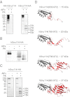
TRiC recognizes and encapsulates C-terminal segments of hSnu114. (A) Western blot analysis of HA-tagged fragments of hSnu114 protease-protected by TRiC encapsulation. HA-hSnu114 and hSnu114-HA were translated in RRL in the absence of radiolabeled amino acid. PK treatment was performed in the presence of ATP·AlFx (see Fig. 3B), followed by SDS/PAGE and Western blotting with anti-HA antibody. Protease-protected fragments of hSnu114-HA are indicated. The positions of proteolytic fragments and molecular weight markers are indicated as in Fig. 4B. (B) Native PAGE analysis of TRiC:hSnu114-HA complexes. The experiment was performed as in Fig. 3C, except that analysis was by anti-HA Western blotting. (C) Anti-HA Western blot analysis of TRiC-bound translation products. TRiC-bound material was excised from native PAGE gels and reanalyzed by SDS/PAGE (see Fig. 3D). (D) Putative structures of hSnu114-HA fragments encapsulated by TRiC. C-terminal proteolytic fragments observed in A and C were mapped on the structural model (Fig. 4A). Note that the hSnu114 sequence contains additional 17 amino acid residues at the C terminus, which were not included in the model. The largest fragment corresponds approximately to domains IV (red) and V (gray) (Fig. 4A).
Fig. 6.
Fig. 6.
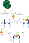
Folding of multidomain proteins by TRiC. Hypothetical model for the folding of a two-domain protein having a TRiC-dependent C-terminal domain, analogous to the GFP-actin fusion protein. Step 1, the N-terminal domain is TRiC-independent and folds cotranslationally, presumably with the aid of ribosome-associated chaperones and the Hsp70 system (59, 70). Step 2, the folding intermediate containing a nonnative C-terminal domain is stabilized against aggregation by Hsp70 or prefoldin and is posttranslationally transferred to TRiC. Alternatively, TRiC may interact cotranslationally with a chaperonin-dependent domain (59, 60). Step 3, the C-terminal domain is encapsulated by TRiC upon ATP hydrolysis and is induced to fold in the specialized physical environment of the chaperonin cavity (23). The iris-like closing mechanism allows the flexible interdomain linker to protrude through the apical pore. Step 4, upon opening of the TRiC cavity, the substrate is either released as successfully folded, native protein or enters another folding cycle.

Similar articles

Cited by

References

    1. Ellis RJ. Protein folding: Importance of the Anfinsen cage. Curr Biol. 2003;13(22):R881–R883. - PubMed
    1. Gershenson A, Gierasch LM. Protein folding in the cell: Challenges and progress. Curr Opin Struct Biol. 2011;21(1):32–41. - PMC - PubMed
    1. Hartl FU. Molecular chaperones in cellular protein folding. Nature. 1996;381(6583):571–579. - PubMed
    1. Horwich AL, Fenton WA, Chapman E, Farr GW. Two families of chaperonin: Physiology and mechanism. Annu Rev Cell Dev Biol. 2007;23:115–145. - PubMed
    1. Frydman J. Folding of newly translated proteins in vivo: The role of molecular chaperones. Annu Rev Biochem. 2001;70:603–647. - PubMed

Publication types

MeSH terms

LinkOut - more resources