. 2012 Jun;1(6):469-79.
doi: 10.5966/sctm.2011-0046. Epub 2012 May 30.

Enhanced lithium-induced brain recovery following cranial irradiation is not impeded by inflammation

Affiliations

Enhanced lithium-induced brain recovery following cranial irradiation is not impeded by inflammation

Jordane Malaterre et al. Stem Cells Transl Med. 2012 Jun.

Abstract

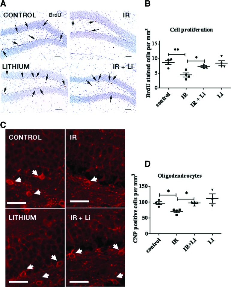
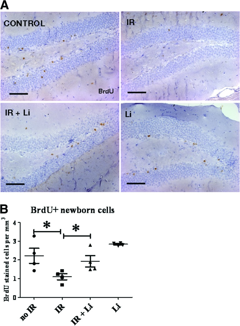
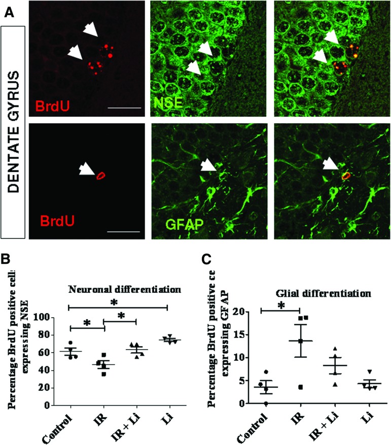
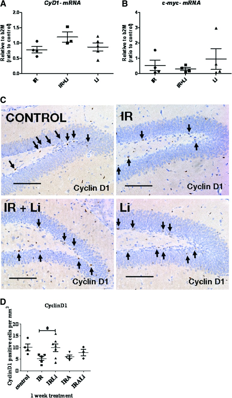
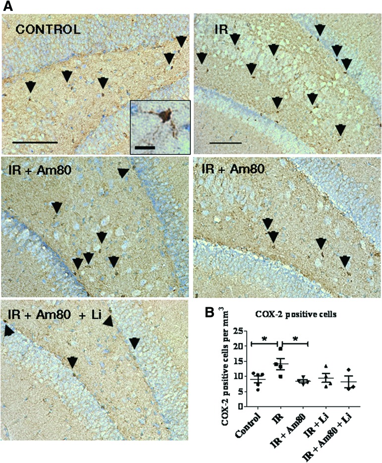
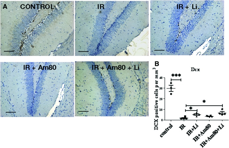
Radiation-induced brain injury occurs in many patients receiving cranial radiation therapy, and these deleterious effects are most profound in younger patients. Impaired neurocognitive functions in both humans and rodents are associated with inflammation, demyelination, and neural stem cell dysfunction. Here we evaluated the utility of lithium and a synthetic retinoid receptor agonist in reducing damage in a model of brain-focused irradiation in juvenile mice. We found that lithium stimulated brain progenitor cell proliferation and differentiation following cranial irradiation while also preventing oligodendrocyte loss in the dentate gyrus of juvenile mice. In response to inflammation induced by radiation, which may have encumbered the optimal reparative action of lithium, we used the anti-inflammatory synthetic retinoid Am80 that is in clinical use in the treatment of acute promyelocytic leukemia. Although Am80 reduced the number of cyclooxygenase-2-positive microglial cells following radiation treatment, it did not enhance lithium-induced neurogenesis recovery, and this alone was not significantly different from the effect of lithium on this proinflammatory response. Similarly, lithium was superior to Am80 in supporting the restoration of new doublecortin-positive neurons following irradiation. These data suggest that lithium is superior in its restorative effects to blocking inflammation alone, at least in the case of Am80. Because lithium has been in routine clinical practice for 60 years, these preclinical studies indicate that this drug might be beneficial in reducing post-therapy late effects in patients receiving cranial radiotherapy and that blocking inflammation in this context may not be as advantageous as previously suggested.


Figures

Figure 1.
Figure 1.
Short-term lithium treatment significantly reverses irradiation-induced decrease in neural stem and progenitor cell (NSPC) proliferation and protects oligodendrocytes in vivo in the dentate gyrus (DG). Experiments were performed using four groups of mice: (a) control, (b) irradiated, (c) lithium-treated, and (d) irradiated and lithium-treated. The mouse groups were fed with control or lithium chow for 1 week starting 2 days after radiation to avoid interfering with cell death and repair. All of the groups were injected with BrdU 1 hour before sacrifice to label NSPCs engaged in the S phase. (A): Hematoxylin-stained coronal sections through the DG showing proliferating BrdU+ cells typically located in the subgranular zone. Lithium increased the number of proliferating cells in the DG, thus reversing irradiation-induced loss of BrdU+ cells. (B): BrdU+ cell quantitation showed that lithium induced a significant increase in the number of BrdU+ cells following irradiation compared with irradiated mice. (C): Oligodendrocytes were identified by immunohistochemical staining for CNP. Typical CNP staining was observed in oligodendrocytes processes and around the cell bodies. Irradiation reduced the number CNP+ cell bodies in the DG (white arrows), whereas lithium blocked this effect. Scale bars = 100 μm. (D): Quantitation of the number of CNP+ cells showed that irradiation produced a significant reduction in the number of oligodendrocytes, whereas lithium restored their numbers to control levels. The data shown are the means ± SEM. *, p < .05. Abbreviations: BrdU, 5-bromo-2′-deoxyuridine; IR, irradiation; CNP, 2′,3′-cyclic nucleotide-3′-phosphohydrolase.
Figure 2.
Figure 2.
A single dose of 8 Gy produces subtle differences in learning and memory ability as assessed using the Morris water maze (MWM). We used the open-field paradigm to assess locomotor and exploratory activity prior to testing the mice in the MWM. (A, B): The mice were first tested in a small open field, and their locomotor behavior was monitored over 3 successive days. A robust habituation was observed in both irradiated and control mice, 4 (A) and 8 (B) weeks after radiation. Hippocampus-related learning and memory function was assessed using the MWM. During five training sessions, a significantly decreased latency to find the platform across sessions was found in both irradiated and control groups 4 (C) and 8 (D) weeks after radiation. The mice were subsequently reassessed for spatial learning to determine apparent neural liability in target reacquisition. Subtle differences were observed between group at 4 weeks after radiation (E) (session 5) and 8 weeks after radiation (F) (session 2). The data shown are the means ± SEM. *, p < .05. Abbreviation: IR, irradiation.
Figure 3.
Figure 3.
Lithium increases the number of newborn cells in the dentate gyrus (DG) following brain-focused irradiation. The mice received BrdU injections twice daily for 3 days 1 week after radiation to label newly generated cells, and the number of surviving newborn cells was assessed after 3 additional weeks. The mice were fed with lithium or control chow for a total of 4 weeks. (A): Newly generated cells were detected by immunohistochemistry for BrdU on coronal sections through the DG. BrdU+ cells were typically seen in the granular layer, in the hilus, and occasionally in the molecular layer of the DG. Irradiation induced a reduction of the number of BrdU+ cell, and lithium reversed this effect. (B): BrdU+ quantitation showed that irradiation produced a 50% decrease in the number of newborn cells after 3 weeks, whereas lithium induced significant recovery in their number. The data shown are the means ± SEM. Scale bar = 100 μm. *, p < .05. Abbreviations: IR, irradiation; BrdU, 5-bromo-2′-deoxyuridine.
Figure 4.
Figure 4.
Lithium increases the percentage of newborn cells that differentiate into neurons under basal conditions and following irradiation but does not influence glial cell differentiation. (A): To assess newborn cell fate in the dentate gyrus (DG), sections were costained for BrdU and NSE to identify neurons or for BrdU and GFAP to identify astrocytes (white arrows) and were analyzed by confocal microscopy. (B): Results of confocal analyses revealed that irradiation significantly reduced neuronal differentiation, whereas long-term lithium treatment significantly enhanced neuronal differentiation at a basal level and following irradiation in the DG. (C): Irradiation led to an increased production of glia (gliosis), whereas lithium did not have any significant effect on glial differentiation. The data shown are the means ± SEM. Scale bars = 50 μm. *, p < .05; **, p < .01. Abbreviations: BrdU, 5-bromo-2′-deoxyuridine; NSE, neuron-specific enolase; GFAP, glial fibrillary acidic protein; IR, irradiation.
Figure 5.
Figure 5.
Effect of lithium treatment on Cyclin D1 and c-Myc expression following irradiation in vitro and in vivo. (A, B): Cyclin D1 (A) and c-Myc (B) mRNA expression was measured in the hippocampus by quantitative reverse transcription-polymerase chain reaction 1 week following brain-focused irradiation. (C): Immunohistochemistry (IHC) detection of Cyclin D1 was performed on brain sections obtained from control, irradiated, and lithium-treated mice. Photographs taken from stained coronal sections show that Cyclin D1+ cells were more abundant in lithium-treated samples compared with untreated samples following irradiation. Scale bars = 100 μm. (D): Quantitation of IHC for Cyclin D1. Lithium treatment restored the number of Cyclin D1+ cells to control levels following irradiation. Am80 treatment did not change the number of Cyclin D1+ cells. The data shown are the means ± SEM. *, p < .05. Abbreviations: CyD1, Cyclin D1; IR, irradiation; IRA, irradiation and lithium; b2M, β2-microglobulin.
Figure 6.
Figure 6.
Effect of 4 weeks of lithium and Am80 treatment on the number of cyclooxygenase-2-positive (COX-2+) microglial cells in the dentate gyrus (DG) following brain-focused irradiation. (A): Immunohistochemistry for COX-2 was performed to assess the effect of Am80 on brain inflammation following cranial irradiation. Typical COX-2+ microglia cells (as shown in the inset) were seen in the hilus, subgranular, and granular layer of the DG (black arrowheads). (B): Positive COX-2 microglia cells were scored to show that their number was significantly elevated following brain-focused irradiation. Administration of Am80 for 4 weeks significantly blocked this effect. Scale bars = 100 μm and scale bar = 20 μm for inset. The data shown are the means ± SEM. *, p < .05. Abbreviation: IR, irradiation.
Figure 7.
Figure 7.
Effect of lithium and Am80 treatment on neurogenesis following brain-focused irradiation. (A): Immunohistochemistry for DCX, a marker for neuroblasts, was used to assess the effect of drug treatment on neurogenesis. (B): Quantitation showed that DCX+ cells were significantly decreased by cranial irradiation when assessed after 4 weeks, whereas lithium treatment for 4 weeks after radiation significantly increased DCX+ cell number compared with irradiated mice but did not revert to control levels. Am80 did not have any significant effect on DCX+ cell number, and concomitant administration with lithium did not produce any additive effect. Scale bars = 100 μm. The data shown are the means ± SEM. *, p < .05; **, p < .01; ***, p < .001. Abbreviations: IR, irradiation; Dcx or DCX, doublecortin.

Similar articles

Cited by

References

    1. Hertzberg H, Huk WJ, Ueberall MA, et al. CNS late effects after ALL therapy in childhood. Part I: Neuroradiological findings in long-term survivors of childhood ALL—An evaluation of the interferences between morphology and neuropsychological performance. The German Late Effects Working Group. Med Pediatr Oncol. 1997;28:387–400. - PubMed
    1. Riva D, Giorgi C. The neurodevelopmental price of survival in children with malignant brain tumours. Childs Nerv Syst. 2000;16:751–754. - PubMed
    1. Roman DD, Sperduto PW. Neuropsychological effects of cranial radiation: Current knowledge and future directions. Int J Radiat Oncol Biol Phys. 1995;31:983–998. - PubMed
    1. Alvarez-Buylla A, Lim DA. For the long run: Maintaining germinal niches in the adult brain. Neuron. 2004;41:683–686. - PubMed
    1. Doetsch F, Garcia-Verdugo JM, Alvarez-Buylla A. Cellular composition and three-dimensional organization of the subventricular germinal zone in the adult mammalian brain. J Neurosci. 1997;17:5046–5061. - PMC - PubMed

Publication types

MeSH terms