Review
. 2012 Jun;1(6):480-91.
doi: 10.5966/sctm.2011-0056. Epub 2012 May 30.

Concise review: the periosteum: tapping into a reservoir of clinically useful progenitor cells

Affiliations
Review

Concise review: the periosteum: tapping into a reservoir of clinically useful progenitor cells

Hana Chang et al. Stem Cells Transl Med. 2012 Jun.

Abstract

Elucidation of the periosteum and its regenerative potential has become a hot topic in orthopedics. Yet few review articles address the unique features of periosteum-derived cells, particularly in light of translational therapies and engineering solutions inspired by the periosteum's remarkable regenerative capacity. This review strives to define periosteum-derived cells in light of cumulative research in the field; in addition, it addresses clinical translation of current insights, hurdles to advancement, and open questions in the field. First, we examine the periosteal niche and its inhabitant cells and the key characteristics of these cells in the context of mesenchymal stem cells and their relevance for clinical translation. We compare periosteum-derived cells with those derived from the marrow niche in in vivo studies, addressing commonalities as well as features unique to periosteum cells that make them potentially ideal candidates for clinical application. Thereafter, we review the differentiation and tissue-building properties of periosteum cells in vitro, evaluating their efficacy in comparison with marrow-derived cells. Finally, we address a new concept of banking periosteum and periosteum-derived cells as a novel alternative to currently available autogenic umbilical blood and perinatal tissue sources of stem cells for today's population of aging adults who were "born too early" to bank their own perinatal tissues. Elucidating similarities and differences inherent to multipotent cells from distinct tissue niches and their differentiation and tissue regeneration capacities will facilitate the use of such cells and their translation to regenerative medicine.


Figures

Figure 1.
Figure 1.
Experimental model elucidating the regenerative capacity of periosteum to regenerate bone in a critical-sized long bone defect. (A): A one-stage bone transport procedure was used, carefully peeling back the periosteum from the bone proximal to the critical-sized defect, osteotomizing, transporting, and docking the denuded bone in the distal defect space, creating a new defect, which was enveloped by the in situ periosteum sleeve (sutured back in place). (B): High-resolution microcomputed tomographs (microCT) showed no bridging of the untreated critical-sized defect (baseline control, group 1) and complete bridging of the defects treated with periosteum. (C): Quantitative density measurements from microCT showed no differences in quantity of tissue generated within the defects treated with periosteum (groups 2–5, data not shown), albeit significant differences are shown in the density of bone and callus, with superior density observed in group 4, followed by group 5. (D): The experimental design allowed for treatment with periosteum in situ in all experimental groups (2–5), with addition of morcellized graft from the iliac crest (groups 3 and 5) as well as retention of small, vascularized bone chips on the inner surface of the periosteum (groups 4 and 5). Adapted from [10]; used with permission of the Journal of Bone and Joint Surgery (http://jbjs.org). Abbreviation: MG/CCM: milligrams per cubic centimeter.
Figure 2.
Figure 2.
Schematic representation (left) and light micrograph (right) depicting the three zones of the periosteum as well as the distribution of cell populations (fibroblasts, pericytes, stem cells, and osteoblasts) and extracellular matrix fibers (Sharpey's fibers and collagen) that contribute to the biological and mechanical properties of the periosteum. Light micrograph image from [17], used with permission.
Figure 3.
Figure 3.
Chondrogenic capacity of periosteum-derived cells (PDCs) is retained with age. PDCs were cultured with or without TGF-β1 for 6 days, fixed, and stained with Alcian Blue. Samples are labeled with the ages of the donors, and asterisks indicate periosteum samples obtained post mortem. After [58]; used with permission. Abbreviation: TGF-β1, transforming growth factor-β1.
Figure 4.
Figure 4.
An overview of the animals (ordered by size), source (ordered by bone type), and age of periosteum-derived cells used in a cross-section of major published studies. Data points indicate reference numbers. Further information is found in supplemental online Table 1.
Figure 5.
Figure 5.
An overview of typical periosteum-derived cell isolation protocols, including harvesting of periosteum cells, mincing of periosteum tissue, and extraction of cells via migration (top) or chemical digestion (bottom). Abbreviations: dPDC, chemically digested periosteum-derived cell; mPDC, migrated periosteum-derived cell.
Figure 6.
Figure 6.
An overview of the harvest methods, isolation protocols, and duration of culture for a cross-section of published studies using periosteum-derived cells. Data points indicate reference numbers. Further information is found in supplemental online Table 2.
Figure 7.
Figure 7.
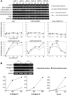
Periosteum-derived cells indicate more osteogenic differentiation on machined titanium surfaces (A), and bone marrow-derived multipotent mesenchymal stromal cells indicate more osteogenic differentiation on acid-etched titanium surfaces (B). Shown is the expression of bone extracellular matrix-related genes analyzed by polymerase chain reaction analysis visualized with ethidium bromide staining and by reverse transcription-polymerase chain reaction for the expression time course for each gene. Genes are normalized to GAPDH expression. Bars indicate SD. Adapted from [74, 120]; used with permission. Abbreviations: AE, acid-etched surface; ALP, alkaline phosphatase; GAPDH, glyceraldehyde-3-phosphate dehydrogenase; M, machined surface.

References

    1. Duhamel HL. Sur le Developpement et la Crue des os des animaux. Mem Acad Roy Des Sci. 1742;55:354–357.
    1. Ollier L. Recherches experimentales sur les greffes osseuses. J Physiol Homme Animaux. 1860;3:88.
    1. Ueno T, Kagawa T, Mizukawa N, et al. Cellular origin of endochondral ossification from grafted periosteum. Anat Rec. 2001;264:348–357. - PubMed
    1. Young RW. Cell proliferation and specialization during endochondral osteogenesis in young rats. J Cell Biol. 1962;14:357–370. - PMC - PubMed
    1. Yu YY, Lieu S, Lu C, et al. Bone morphogenetic protein 2 stimulates endochondral ossification by regulating periosteal cell fate during bone repair. Bone. 2010;47:65–73. - PMC - PubMed

Publication types