. 2012 Jun;1(6):510-9.
doi: 10.5966/sctm.2012-0002. Epub 2012 Jun 11.

Perivascular stem cells: a prospectively purified mesenchymal stem cell population for bone tissue engineering

Affiliations

Perivascular stem cells: a prospectively purified mesenchymal stem cell population for bone tissue engineering

Aaron W James et al. Stem Cells Transl Med. 2012 Jun.

Abstract

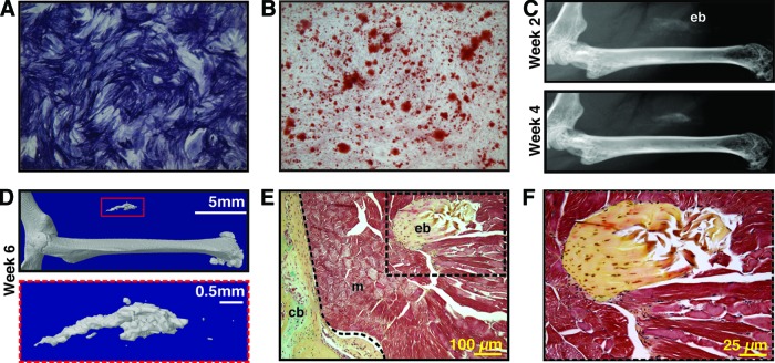
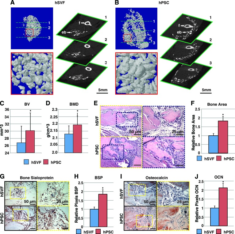
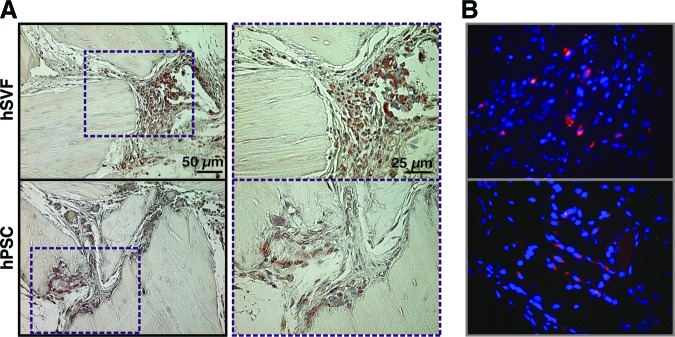
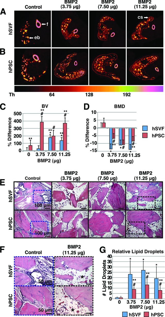
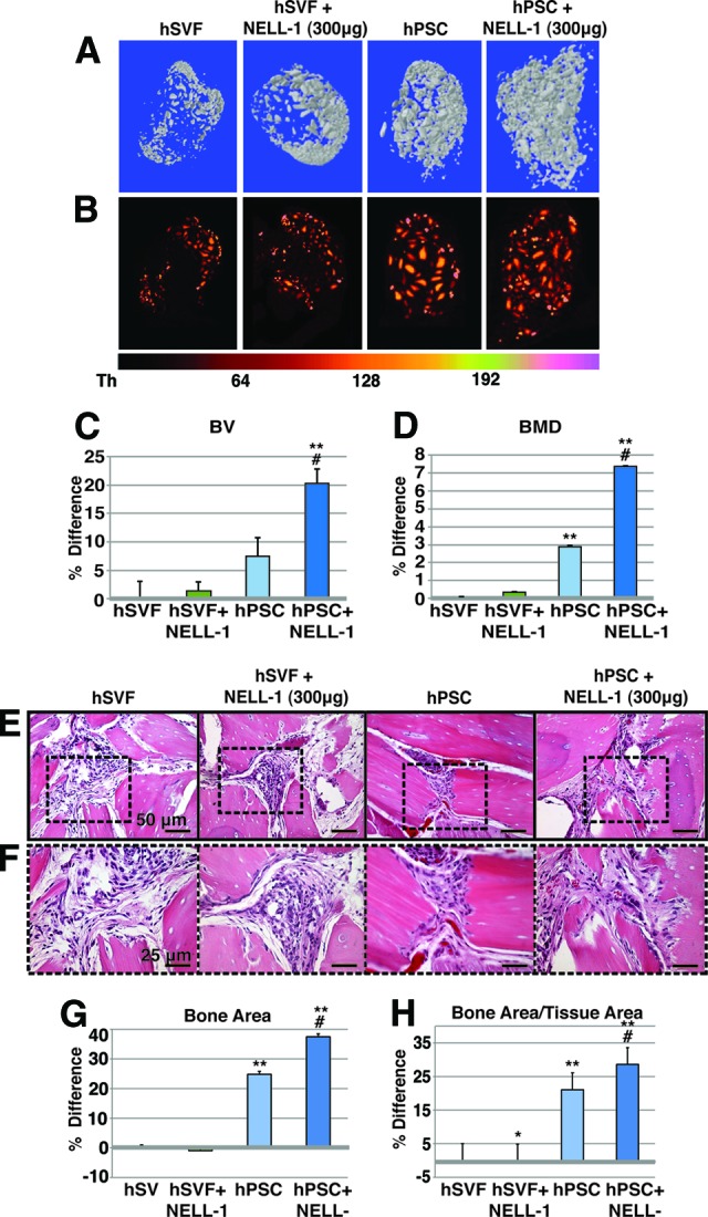
Adipose tissue is an ideal source of mesenchymal stem cells for bone tissue engineering: it is largely dispensable and readily accessible with minimal morbidity. However, the stromal vascular fraction (SVF) of adipose tissue is a heterogeneous cell population, which leads to unreliable bone formation. In the present study, we prospectively purified human perivascular stem cells (PSCs) from adipose tissue and compared their bone-forming capacity with that of traditionally derived SVF. PSCs are a population (sorted by fluorescence-activated cell sorting) of pericytes (CD146+CD34-CD45-) and adventitial cells (CD146-CD34+CD45-), each of which we have previously reported to have properties of mesenchymal stem cells. Here, we found that PSCs underwent osteogenic differentiation in vitro and formed bone after intramuscular implantation without the need for predifferentiation. We next sought to optimize PSCs for in vivo bone formation, adopting a demineralized bone matrix for osteoinduction and tricalcium phosphate particle formulation for protein release. Patient-matched, purified PSCs formed significantly more bone in comparison with traditionally derived SVF by all parameters. Recombinant bone morphogenetic protein 2 increased in vivo bone formation but with a massive adipogenic response. In contrast, recombinant Nel-like molecule 1 (NELL-1; a novel osteoinductive growth factor) selectively enhanced bone formation. These studies suggest that adipose-derived human PSCs are a new cell source for future efforts in skeletal regenerative medicine. Moreover, PSCs are a stem cell-based therapeutic that is readily approvable by the U.S. Food and Drug Administration, with potentially increased safety, purity, identity, potency, and efficacy. Finally, NELL-1 is a candidate growth factor able to induce human PSC osteogenesis.


Figures

Figure 1.
Figure 1.
Human perivascular stem cells (hPSCs) are an osteocompetent cell population. (A, B): hPSCs were cultured under osteogenic conditions (10% fetal bovine serum, 100 μg/ml ascorbic acid, 10 mM β-glycerophosphate). (A): Representative image of alkaline phosphatase staining at 5 days of differentiation. (B): Representative image of alizarin red staining, 10 days of differentiation. (C–F): hPSCs were implanted in the thigh complex of a SCID mouse using a collagen sponge carrier (2.5 × 10⁵ cells, sponge size 2.0 × 1.0 × 0.5 cm). (C): High-resolution radiography at 2 and 4 weeks postimplantation. (D): Three-dimensional microcomputed tomography reconstructions at threshold 90. (E, F): Pentachrome staining of histological sections. n = 5 implants, from n = 1 patient specimens. See supplemental online Table 2 for treatment groups. Abbreviations: cb, cortical bone; eb, ectopic bone; m, muscle.
Figure 2.
Figure 2.
hPSCs undergo more robust differentiation in comparison with hSVF. Equal numbers of viable hSVF cells or hPSCs (2.5 × 10⁵) from the same patient samples were implanted intramuscularly in the thigh of a SCID mouse. An osteoinductive DBX Putty was used as scaffold. Assessments were performed at 4 weeks postimplantation. (A, B): Microcomputed tomography (microCT) images of hSVF- and hPSC-treated samples. Representative three-dimensional microCT reconstructions at threshold (Th) 90 (left) and corresponding two-dimensional axial slices (right). (C, D): Analysis of BV and BMD among hSVF- and hPSC-treated samples; Th50–120. (E): Representative hematoxylin and eosin staining. (F): Representative histomorphometric quantification of bone area per ×100 field. (G): Representative BSP immunohistochemistry. (H): Quantification of relative staining per ×400 field. (I): Representative OCN immunohistochemistry. (J): Quantification of relative staining per ×400 field. Histomorphometric quantification calculated from n = 6 random microscopical fields. n = 12 implants per cell type, n = 3 patient specimens. See supplemental online Table 3 for treatment groups. *, p < .05 in comparison with control as assessed by Student's t test. Abbreviations: BMD, bone mineral density; BSP, bone sialoprotein; BV, bone volume; eb, ectopic bone; f, femur; hPSC, human perivascular stem cell; hSVF, human stromal vascular fraction; OCN, osteocalcin.
Figure 3.
Figure 3.
Persistence of hSVF or hPSCs after implantation. Shown are hSVF- or hPSC-treated samples implanted intramuscularly on a DBX Putty scaffold and assessed for cell persistence 4 weeks postimplantation. (A): Human major histocompatibility complex class I immunohistochemistry, indicating the presence of human xenografted cells. (B): Fluorescent cell labeling (PKH) performed prior to implantation. Red indicates persistence of human cells, and Hoechst nuclear counterstain appears blue. Abbreviations: hPSC, human perivascular stem cell; hSVF, human stromal vascular fraction.
Figure 4.
Figure 4.
Effects of BMP2 on hSVF- or hPSC-mediated ectopic bone formation. hSVF or hPSCs were implanted intramuscularly with or without BMP2 on a DBX Putty (DBX) scaffold and assessed 4 weeks postimplantation. See supplemental online Table 5 for treatment groups. (A, B): Colorized representative two-dimensional axial microcomputed tomography images. A scale bar below indicates colorization at various thresholds. (C): Relative BV. Values are normalized to hSVF + DBX only. Threshold 100 was used. (D): Relative BMD. Values are normalized to hSVF + DBX only. (E, F): Representative hematoxylin and eosin images at low (E) and high (F) magnifications. n = 8 implants per cell type, n = 1 patient specimen. (G): Relative lipid droplet number per ×100 field. Means were calculated from 10 random microscopic fields. *, p < .05 in comparison with hSVF control; #, p < .05 in comparison with hPSC control; **, p < .05 in comparison with hSVF with same dose of BMP2. See supplemental online Table 4 for treatment groups. Abbreviations: BMD, bone mineral density; BMP, bone morphogenetic protein; BV, bone volume; cs, cortical shell; eb, ectopic bone; f, femur; hPSC, human perivascular stem cell; hSVF, human stromal vascular fraction.
Figure 5.
Figure 5.
Effects of NELL-1 on hPSC- or hSVF-mediated ectopic bone formation. hSVF or hPSCs in a SCID mouse muscle pouch with DBX Putty, with or without NELL-1, assessed at 4 weeks postimplantation. See supplemental online Table 6 for treatment groups. (A): Representative three-dimensional microcomputed tomography (microCT) reconstructions. Threshold (Th) 90 was used. (B): Colorized representative two-dimensional microCT images. A scale bar below indicates colorization at various thresholds. (C): Relative BV; Th50–120 used. (D): Relative BMD. (E, F): Representative hematoxylin and eosin staining at low (E) and high (F) magnifications. (G, H): Histomorphometric analysis of mean bone area (G) and mean bone area/tissue area (H), as calculated from 30 random high-powered-fields. n = 9 implants per treatment group, n = 1 patient specimen. *, p < .05 in comparison with hSVF control; #, p < .05 in comparison with hPSC control; **, p < .05 in comparison with hSVF with same dose NELL-1. See supplemental online Table 5 for treatment groups. Abbreviations: BMD, bone mineral density; BV, bone volume; hPSC, human perivascular stem cell; hSVF, human stromal vascular fraction; NELL-1, Nel-like molecule 1.
Figure 6.
Figure 6.
Human white adipose tissue houses distinct pericyte and adventitial cell populations, which together represent perivascular stem cells (PSCs). (A, B): After exclusion of DAPI+ dead cells (A) and CD45+ hematopoietic cells (B), two PSC populations were isolated with differential expression of CD34 and CD146. (C): Distinct CD34+CD146− adventitial cells and CD34−CD146+ pericytes were obtained and combined for in vivo implantation. (D): The relative numbers of pericytes and adventitial cells are depicted as a fraction of the total DAPI− SVF cells. Pericytes and adventitial cells make up approximately 17.1% and 22.5%, respectively. PSCs then represent on average 39.6% of total viable cells. Abbreviations: DAPI, 4′,6-diamidino-2-phenylindole; FSC, forward scatter; SSC, side scatter; SVF, stromal vascular fraction.

References

    1. Zuk PA, Zhu M, Ashjian P, et al. Human adipose tissue is a source of multipotent stem cells. Mol Biol Cell. 2002;13:4279–4295. - PMC - PubMed
    1. Zuk PA. The adipose-derived stem cell: Looking back and looking ahead. Mol Biol Cell. 2010;21:1783–1787. - PMC - PubMed
    1. Laurencin C, Khan Y. Bone Graft Substitute Materials. emedicine 2004. [Accessed February 16, 2012]. Available at http://emedicine.medscape.com/article/1230616-overview.
    1. Laurie SW, Kaban LB, Mulliken JB, et al. Donor-site morbidity after harvesting rib and iliac bone. Plast Reconstr Surg. 1984;73:933–938. - PubMed
    1. Frodel JL, Jr., Marentette LJ, Quatela VC, et al. Calvarial bone graft harvest. Techniques, considerations, and morbidity. Arch Otolaryngol Head Neck Surg. 1993;119:17–23. - PubMed

Publication types

MeSH terms