. 2012 Sep;1(9):641-50.
doi: 10.5966/sctm.2012-0043. Epub 2012 Sep 7.

Variability in the generation of induced pluripotent stem cells: importance for disease modeling

Affiliations

Variability in the generation of induced pluripotent stem cells: importance for disease modeling

Alejandra M Vitale et al. Stem Cells Transl Med. 2012 Sep.

Abstract

In the field of disease modeling, induced pluripotent stem cells (iPSCs) have become an appealing choice, especially for diseases that do not have an animal model. They can be generated from patients with known clinical features and compared with cells from healthy controls to identify the biological bases of disease. This study was undertaken to determine the variability in iPSC lines derived from different individuals, with the aim of determining criteria for selecting iPSC lines for disease models. We generated and characterized 18 iPSC lines from eight donors and considered variability at three levels: (a) variability in the criteria that define iPSC lines as pluripotent cells, (b) variability in cell lines from different donors, and (c) variability in cell lines from the same donor. We found that variability in transgene expression and pluripotency marker levels did not prevent iPSCs from fulfilling all other criteria for pluripotency, including teratoma formation. We found low interindividual and interclonal variability in iPSCs that fulfilled the most stringent criteria for pluripotency, with very high correlation in their gene expression profiles. Interestingly, some cell lines exhibited reprogramming instability, spontaneously regressing from a fully to a partially reprogrammed state. This was associated with a low percentage of cells expressing the pluripotency marker stage-specific embryonic antigen-4. Our study shows that it is possible to define a similar "ground state" for each cell line as the basis for making patient versus control comparisons, an essential step in order to identify disease-associated variability above individual and cell line variability.


Figures

Figure 1.
Figure 1.
Characterization of induced pluripotent stem cell (iPSC) lines: representative pictures. (A): Polymerase chain reaction analysis of endogenous pluripotency markers SOX2 (151 bp), KLF4 (397 bp; lower band corresponds to primer dimers), OCT4 (144 bp), and GAPDH (94 bp). (B): Phase image and SSEA4/DAPI staining of iPSC colony. Magnification, ×40. (C): Normal karyotype. (D): Hematoxylin and eosin staining of a teratoma showing differentiation into ectoderm, mesoderm, and endoderm. (E): Flow cytometry analysis for pluripotency markers OCT4, SOX2, NANOG, SSEA4, and TRA1-60. Abbreviations: DAPI, 4′,6-diamidino-2-phenylindole; GAPDH, glyceraldehyde-3-phosphate dehydrogenase; SSEA4, stage-specific embryonic antigen-4.
Figure 2.
Figure 2.
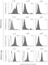
Flow cytometry analysis of SSEA4 protein expression. SSEA4 protein levels were quantified at three different passages in three SSEA4-low induced pluripotent stem cell (iPSC) lines and one representative SSEA4-high iPSC line. The percentages of cells expressing the marker (gray) above the isotype-control cells (clear) are shown in the graph. *, Cells sorted for transcriptome analysis. Abbreviation: SSEA4, stage-specific embryonic antigen-4.
Figure 3.
Figure 3.
Principal component analysis shows gene expression variability among different clones and cell types. Squares, fibroblasts; triangles, iPSC SSEA4-low; circles, iPSC SSEA4-high; diamonds, hESCs. Symbols of the same color indicate the same patient. Abbreviations: hESC, human embryonic stem cell; iPSC, induced pluripotent stem cell; PCA, principal component analysis; SSEA4, stage-specific embryonic antigen-4.
Figure 4.
Figure 4.
Gene expression levels of pluripotency markers (OCT4, NANOG, SOX2, cMYC, and KLF4) showing variability among different cell lines and cell types. Squares, fibroblasts; triangles, iPSC SSEA4-low; circles, iPSC SSEA4-high; diamonds, hESCs. Abbreviations: hESC, human embryonic stem cell; iPSC, induced pluripotent stem cell; SSEA4, stage-specific embryonic antigen-4.
Figure 5.
Figure 5.
PluriTest output. The background encodes an empirical density map indicating pluripotency and novelty as indicated by the color bar. In the red area, corresponding to fully reprogrammed cells, cluster the iPSC SSEA4-high (yellow circles), hESCs (red circles), and iPSC data provided by Müller et al. [17] (green circles). In the middle area, corresponding to partially reprogrammed cells, are the iPSC SSEA4-low (dark blue circles). Fibroblasts (light blue circles) and astrocytes (purple circle) cluster together at the area corresponding to somatic cells. Circles surrounded by a black line are the results of cell lines provided by Müller et al. [17]. Abbreviations: ESC, embryonic stem cell; iPSC, induced pluripotent stem cell; PSC, pluripotent stem cell; SSEA4, stage-specific embryonic antigen-4.
Figure 6.
Figure 6.
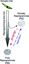
Diagram representing different outcomes of reprogramming progression for the generation of iPSC lines. The success of generating iPSCs depends on the appropriate progression through the reprogramming path from a somatic cell to the fully reprogrammed iPSC. Some cells will deviate from this path, reaching only a partially reprogrammed state. However, these cells may convert subsequently to the fully reprogrammed state. Our results demonstrated the converse, that fully reprogrammed iPSCs can regress to the partially reprogrammed state, even after previously fulfilling stringent definitions of pluripotency, including teratoma formation. Abbreviation: iPSC, induced pluripotent stem cell.

Similar articles

Cited by

References

    1. Vitale AM, Wolvetang E, Mackay-Sim A. Induced pluripotent stem cells: A new technology to study human diseases. Int J Biochem Cell Biol. 2011;43:843–846. - PubMed
    1. Matigian NA, McCurdy RD, Feron F, et al. Fibroblast and lymphoblast gene expression profiles in schizophrenia: Are non-neural cells informative? PLoS One. 2008;3:e2412. - PMC - PubMed
    1. Frumkin T, Malcov M, Telias M, et al. Human embryonic stem cells carrying mutations for severe genetic disorders. In Vitro Cell Dev Biol Anim. 2010;46:327–336. - PubMed
    1. Pickering SJ, Minger SL, Patel M, et al. Generation of a human embryonic stem cell line encoding the cystic fibrosis mutation deltaF508, using preimplantation genetic diagnosis. Reprod Biomed Online. 2005;10:390–397. - PubMed
    1. Park IH, Arora N, Huo H, et al. Disease-specific induced pluripotent stem cells. Cell. 2008;134:877–886. - PMC - PubMed

Publication types