. 2012 Sep;1(9):673-84.
doi: 10.5966/sctm.2012-0053. Epub 2012 Sep 5.

An abundant perivascular source of stem cells for bone tissue engineering

Affiliations

An abundant perivascular source of stem cells for bone tissue engineering

Aaron W James et al. Stem Cells Transl Med. 2012 Sep.

Abstract

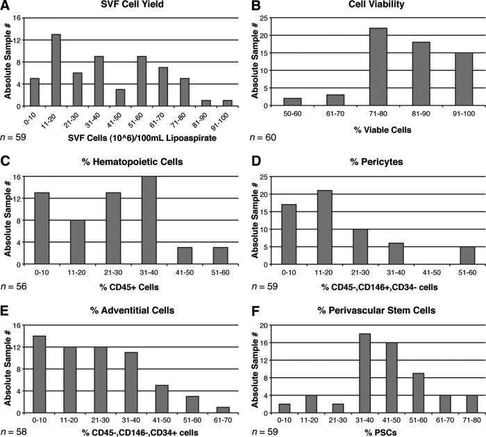
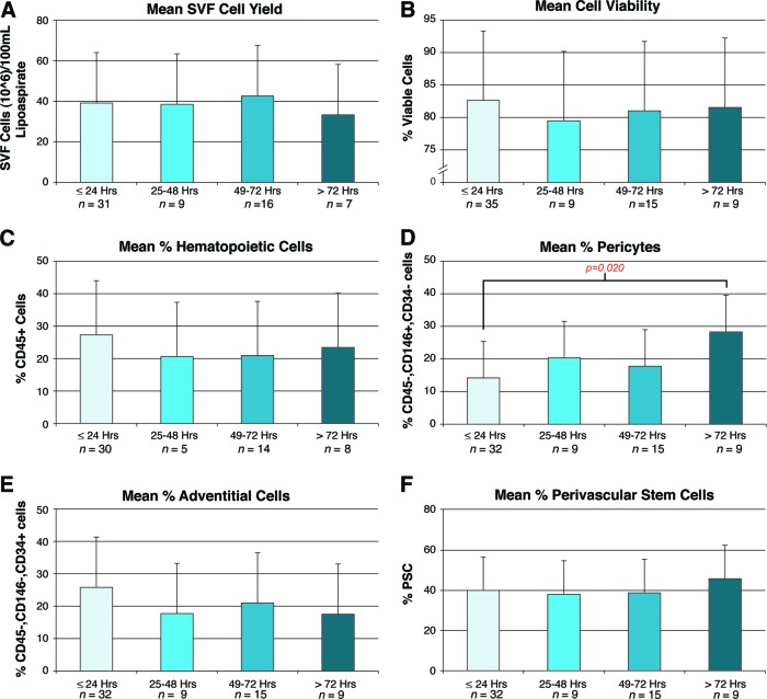
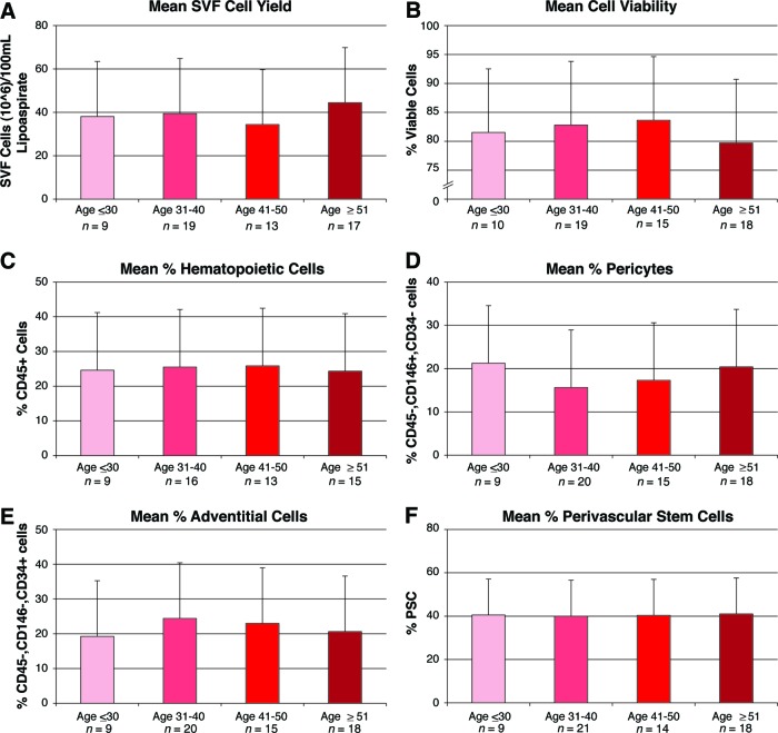
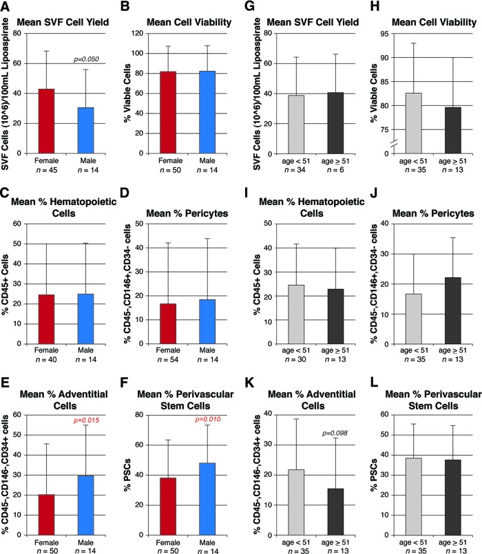
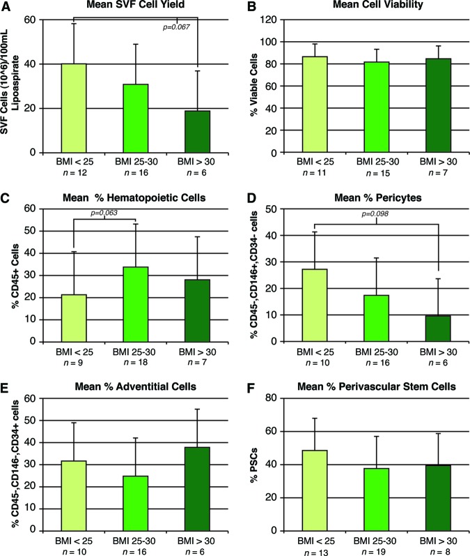
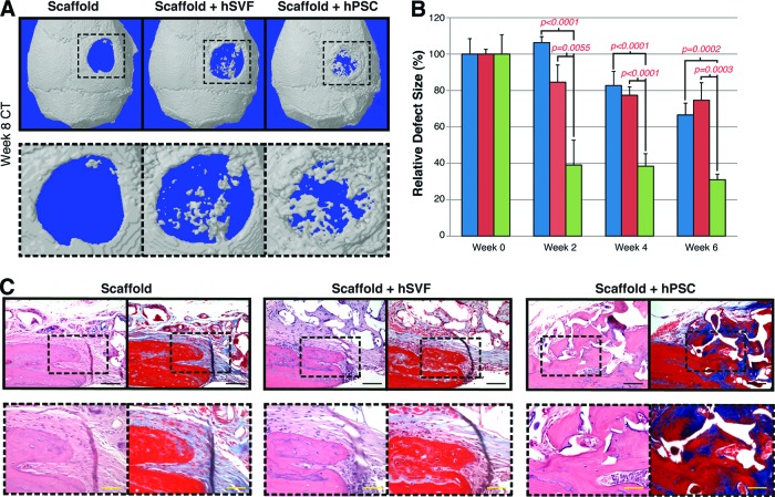
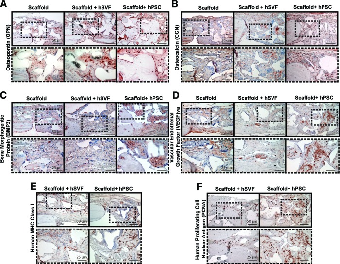
Adipose tissue is an ideal mesenchymal stem cell (MSC) source, as it is dispensable and accessible with minimal morbidity. However, the stromal vascular fraction (SVF) of adipose tissue is a heterogeneous cell population, which has disadvantages for tissue regeneration. In the present study, we prospectively purified human perivascular stem cells (PSCs) from n = 60 samples of human lipoaspirate and documented their frequency, viability, and variation with patient demographics. PSCs are a fluorescence-activated cell sorting-sorted population composed of pericytes (CD45-, CD146+, CD34-) and adventitial cells (CD45-, CD146-, CD34+), each of which we have previously reported to have properties of MSCs. Here, we found that PSCs make up, on average, 43.2% of SVF from human lipoaspirate (19.5% pericytes and 23.8% adventitial cells). These numbers were minimally changed by age, gender, or body mass index of the patient or by length of refrigerated storage time between liposuction and processing. In a previous publication, we observed that human PSCs (hPSCs) formed significantly more bone in vivo in comparison with unsorted human SVF (hSVF) in an intramuscular implantation model. We now extend this finding to a bone injury model, observing that purified hPSCs led to significantly greater healing of mouse critical-size calvarial defects than hSVF (60.9% healing as opposed to 15.4% healing at 2 weeks postoperative by microcomputed tomography analysis). These studies suggest that adipose-derived hPSCs are a new cell source for future efforts in skeletal regenerative medicine. Moreover, hPSCs are a stem cell-based therapeutic that is readily approvable by the U.S. Food and Drug Administration, with potentially increased safety, purity, identity, potency, and efficacy.


Figures

Figure 1.
Figure 1.
Yield, viability, and frequency of human PSCs in human white adipose tissue. (A): Total SVF cell yield, as represented by total SVF cells (106) per 100 ml of tissue lipoaspirate. Blood and saline of lipoaspirate fluid are not included in these calculations. Each bar indicates the number of lipoaspirate samples falling within the stated range of values. (B): Percentage of cell viability, as determined by percentage of DAPI− cells by flow cytometry. (C): Percentage of hematopoietic cells, as determined by percentage of CD45+ cells among viable (DAPI−) cells by flow cytometry. (D): Percentage of pericytes, as determined by percentage of CD146+, CD34− cells among viable (DAPI−) cells. (E): Percentage of adventitial cells, as determined by percentage of CD146−, CD34+ cells among viable (DAPI−) cells. (F): Percentage of human PSCs, as determined by percentage of pericytes + percentage of adventitial cells. Abbreviations: PSC, perivascular stem cell; SVF, stromal vascular fraction.
Figure 2.
Figure 2.
Effects of storage on human PSC yield, viability, and frequency. Samples were shipped and stored as unprocessed lipoaspirate at 4°C prior to digestion, staining, and fluorescence-activated cell sorting isolation. Characteristics of cell yield, viability, and frequency were assessed, stratified by hours of storage. Ranges of hours of storage were 0–24, 25–48, 49–72, and >72 hours, calculated from shipment to processing. Maximum storage time was 168 hours. (A): Total SVF cell yield, as represented by total SVF cells (106) per 100 ml of tissue lipoaspirate. (B): Percentage of cell viability, as determined by percentage of DAPI− cells. (C): Percentage of hematopoietic cells, as determined by percentage of CD45+ cells among DAPI− cells. (D): Percentage of pericytes, as determined by percentage of CD146+, CD34− cells among DAPI− cells. (E): Percentage of adventitial cells, as determined by percentage of CD146−, CD34+ cells among DAPI− cells. (F): Percentage of human PSCs, as determined by percentage of pericytes + percentage of adventitial cells. Abbreviations: PSC, perivascular stem cell; SVF, stromal vascular fraction.
Figure 3.
Figure 3.
Effects of age on human PSC yield, viability, and frequency. Samples were derived from both genders and a wide range of ages (24–69 years). Characteristics of cell yield, viability, and frequency were assessed, stratified by age. Parameters of yield, viability, and frequency were as follows. (A): Total SVF cell yield, as represented by total SVF cells (106) per 100 ml of tissue lipoaspirate. (B): Percentage of cell viability, as determined by percentage of DAPI− cells. (C): Percentage of hematopoietic cells, as determined by percentage of CD45+ cells among DAPI− cells. (D): Percentage of pericytes, as determined by percentage of CD146+, CD34− cells among DAPI− cells. (E): Percentage of adventitial cells, as determined by percentage of CD146−, CD34+ cells among DAPI− cells. (F): Percentage of human PSCs, as determined by percentage of pericytes + percentage of adventitial cells. Abbreviations: PSC, perivascular stem cell; SVF, stromal vascular fraction.
Figure 4.
Figure 4.
Effects of gender on human perivascular stem cell (hPSC) yield, viability, and frequency. Samples were derived from both genders and a wide range of ages (24–69 years). Characteristics of cell yield, viability, and frequency were assessed, stratified by either gender (A–F) or menopausal status (G–L). Menopausal status was estimated on the basis of female age less than or greater than 51. Parameters of yield, viability, and frequency were as follows. (A, G): Total SVF cell yield, as represented by total SVF cells (106) per 100 ml of tissue lipoaspirate. (B, H): Percentage of cell viability, as determined by percentage of DAPI− cells. (C, I): Percentage of hematopoietic cells, as determined by percentage of CD45+ cells among DAPI− cells. (D, J): Percentage of pericytes, as determined by percentage of CD146+, CD34− cells among DAPI− cells. (E, K): Percentage of adventitial cells, as determined by percentage of CD146−, CD34+ cells among DAPI− cells. (F, L): Percentage of hPSCs, as determined by percentage of pericytes + percentage of adventitial cells. Abbreviations: PSC, perivascular stem cell; SVF, stromal vascular fraction.
Figure 5.
Figure 5.
Effects of BMI on human perivascular stem cell (hPSC) yield, viability, and frequency. Samples were derived from nonoverweight (BMI <25 kg/m2), overweight (BMI 25–30 kg/m2), and obese (BMI >30 kg/m2) patients. Characteristics of cell yield, viability, and frequency were stratified by patient BMI. (A): Total SVF cell yield. (B): Percentage of DAPI− cells. (C): Percentage of CD45+ hematopoietic cells. (D): Percentage of pericytes (CD146+, CD34− cells). (E): Percentage of adventitial cells (CD146−, CD34+ cells). (F): Percentage of hPSCs (pericytes + adventitial cells). Abbreviations: BMI, body mass index; PSC, perivascular stem cell; SVF, stromal vascular fraction.
Figure 6.
Figure 6.
Calvarial healing by microCT and histology. hSVF or hPSCs were used to treat a 3-mm-diameter parietal bone defect in a SCID mouse. A custom-made hydroxyapatite-coated poly(lactic-coglycolic acid) scaffold was used as a carrier. Defects were treated with either an empty scaffold or a scaffold seeded with cells (scaffold + hSVF or scaffold + hPSC). Details of treatment groups are given in supplemental online Table 2. (A): Three-dimensional reconstructions of control, hSVF-treated, or hPSC-treated calvarial defects shown at 8 weeks postoperative. A CT threshold of 40 was used. (B): Relative defect healing as assessed by 0, 2, 4, and 6 weeks postoperative by serial live microCT scans. Relative defect area was calculated using a top-down view of the calvaria using AMIDE software images, followed by Adobe Photoshop quantification of relative defect size. (C): Representative hematoxylin/eosin and Masson's trichrome images for the defect site. Images are taken from the lateral defect edge to delineate old from new bone. n = 16–18 mice per treatment group split equally among n = 4 separate patient samples. Patient demographics are given in supplemental online Table 1. Black scale bars = 50 μm. Yellow scale bars = 25 μm. Abbreviations: CT, computed tomography; hPSC, human perivascular stem cell; hSVF, human stromal vascular fraction.
Figure 7.
Figure 7.
Calvarial healing by immunohistochemistry. hSVF or hPSCs were used to treat a 3-mm-diameter parietal bone defect in a SCID mouse. A custom-made hydroxyapatite-coated poly(lactic-coglycolic acid) scaffold was used as a carrier. Defects were treated either with an empty scaffold or with a scaffold seeded with hSVF or hPSCs. Treatment groups are given in supplemental online Table 2. Immunostaining was performed for OPN (A), OCN (B), BMP2 (C), VEGF (D), MHC class I (E), and PCNA (F). n = 16–18 mice per treatment group split equally among n = 4 separate patient samples. Scale bars = 50 μm (top rows), 25 μm (bottom rows). Abbreviations: BMP2, bone morphogenetic protein 2; hPSC, human perivascular stem cell; hSVF, human stromal vascular fraction; MHC, major histocompatibility complex; OCN, osteocalcin; OPN, osteopontin; PCNA, proliferating cell nuclear antigen; VEGF, vascular endothelial growth factor.

References

    1. Levi B, James AW, Nelson ER, et al. Human adipose derived stromal cells heal critical size mouse calvarial defects. PLoS One. 2010;5:e11177. - PMC - PubMed
    1. Levi B, James AW, Nelson ER, et al. Human adipose-derived stromal cells stimulate autogenous skeletal repair via paracrine hedgehog signaling with calvarial osteoblasts. Stem Cells Dev. 2011;20:243–257. - PMC - PubMed
    1. Cui L, Liu B, Liu G, et al. Repair of cranial bone defects with adipose derived stem cells and coral scaffold in a canine model. Biomaterials. 2007;28:5477–5486. - PubMed
    1. Tapp H, Hanley EN, Jr., Patt JC, et al. Adipose-derived stem cells: Characterization and current application in orthopaedic tissue repair. Exp Biol Med (Maywood) 2009;234:1–9. - PubMed
    1. Safwani WK, Makpol S, Sathapan S, et al. The impact of long-term in vitro expansion on the senescence-associated markers of human adipose-derived stem cells. Appl Biochem Biotechnol. 2012;166:2101–2113. - PubMed

Publication types