Mesenchymal stem cell transplantation improves regional cardiac remodeling following ovine infarction
- PMID: 23197875
- PMCID: PMC3659738
- DOI: 10.5966/sctm.2012-0027
Mesenchymal stem cell transplantation improves regional cardiac remodeling following ovine infarction
Erratum in
- Stem Cells Transl Med. 2013 Aug;2(8):633
Abstract
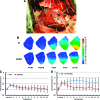
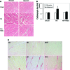
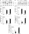
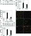
Progressive cardiac remodeling, including the myopathic process in the adjacent zone following myocardial infarction (MI), contributes greatly to the development of cardiac failure. Cardiomyoplasty using bone marrow-derived mesenchymal stem cells (MSCs) has been demonstrated to protect cardiomyocytes and/or repair damaged myocardium, leading to improved cardiac performance, but the therapeutic effects on cardiac remodeling are still under investigation. Here, we tested the hypothesis that MSCs could improve the pathological remodeling of the adjacent myocardium abutting the infarct. Allogeneic ovine MSCs were transplanted into the adjacent zone by intracardiac injection 4 hours after infarction. Results showed that remodeling and contractile strain alteration were reduced in the adjacent zone of the MSC-treated group. Cardiomyocyte hypertrophy was significantly attenuated with the normalization of the hypertrophy-related signaling proteins phosphatidylinositol 3-kinase α (PI3Kα), PI3Kγ, extracellular signal-regulated kinase (ERK), and phosphorylated ERK (p-ERK) in the adjacent zone of the MSC-treated group versus the MI-alone group. Moreover, the imbalance of the calcium-handling proteins sarcoplasmic reticulum Ca(2+) adenosine triphosphatase (SERCA2a), phospholamban (PLB), and sodium/calcium exchanger type 1 (NCX-1) induced by MI was prevented by MSC transplantation, and more strikingly, the activity of SERCA2a and uptake of calcium were improved. In addition, the upregulation of the proapoptotic protein Bcl-xL/Bcl-2-associated death promoter (BAD) was normalized, as was phospho-Akt expression; there was less fibrosis, as revealed by staining for collagen; and the apoptosis of cardiomyocytes was significantly inhibited in the adjacent zone by MSC transplantation. Collectively, these data demonstrate that MSC implantation improved the remodeling in the region adjacent to the infarct after cardiac infarction in the ovine infarction model.
Figures


Similar articles
-
Short-term mechanical unloading with left ventricular assist devices after acute myocardial infarction conserves calcium cycling and improves heart function.JACC Cardiovasc Interv. 2013 Apr;6(4):406-15. doi: 10.1016/j.jcin.2012.12.122. Epub 2013 Mar 20. JACC Cardiovasc Interv. 2013. PMID: 23523452 Free PMC article.
-
AKT-modified autologous intracoronary mesenchymal stem cells prevent remodeling and repair in swine infarcted myocardium.Chin Med J (Engl). 2010 Jul;123(13):1702-8. Chin Med J (Engl). 2010. PMID: 20819633
-
Mesenchymal stem cell therapy associated with endurance exercise training: Effects on the structural and functional remodeling of infarcted rat hearts.J Mol Cell Cardiol. 2016 Jan;90:111-9. doi: 10.1016/j.yjmcc.2015.12.012. Epub 2015 Dec 15. J Mol Cell Cardiol. 2016. PMID: 26705058
-
Exploring Cutting-Edge Approaches to Potentiate Mesenchymal Stem Cell and Exosome Therapy for Myocardial Infarction.J Cardiovasc Transl Res. 2024 Apr;17(2):356-375. doi: 10.1007/s12265-023-10438-x. Epub 2023 Oct 11. J Cardiovasc Transl Res. 2024. PMID: 37819538 Review.
-
Mesenchymal stem cells and the treatment of cardiac disease.Exp Biol Med (Maywood). 2006 Jan;231(1):39-49. doi: 10.1177/153537020623100105. Exp Biol Med (Maywood). 2006. PMID: 16380643 Review.
Cited by
-
MSC Pretreatment for Improved Transplantation Viability Results in Improved Ventricular Function in Infarcted Hearts.Int J Mol Sci. 2022 Jan 8;23(2):694. doi: 10.3390/ijms23020694. Int J Mol Sci. 2022. PMID: 35054878 Free PMC article.
-
miR-10a rejuvenates aged human mesenchymal stem cells and improves heart function after myocardial infarction through KLF4.Stem Cell Res Ther. 2018 May 30;9(1):151. doi: 10.1186/s13287-018-0895-0. Stem Cell Res Ther. 2018. PMID: 29848383 Free PMC article.
-
Comparison of human induced pluripotent stem-cell derived cardiomyocytes with human mesenchymal stem cells following acute myocardial infarction.PLoS One. 2014 Dec 31;9(12):e116281. doi: 10.1371/journal.pone.0116281. eCollection 2014. PLoS One. 2014. PMID: 25551230 Free PMC article.
-
"Second-generation" stem cells for cardiac repair.World J Stem Cells. 2015 Mar 26;7(2):352-67. doi: 10.4252/wjsc.v7.i2.352. World J Stem Cells. 2015. PMID: 25815120 Free PMC article. Review.
-
Effects of transplantation with marrow-derived mesenchymal stem cells modified with survivin on renal ischemia-reperfusion injury in mice.Yonsei Med J. 2014 Jul;55(4):1130-7. doi: 10.3349/ymj.2014.55.4.1130. Yonsei Med J. 2014. PMID: 24954347 Free PMC article.
References
-
- Roe MT, Messenger JC, Weintraub WS, et al. Treatments, trends, and outcomes of acute myocardial infarction and percutaneous coronary intervention. J Am Coll Cardiol. 2010;56:255–262. - PubMed
-
- González A, Ravassa S, Beaumont J, et al. New targets to treat the structural remodeling of the myocardium. J Am Coll Cardiol. 2011;58:1833–1843. - PubMed
-
- Orlic D, Hill JM, Arai AE. Stem Cells for myocardial regeneration. Circ Res. 2002;91:1092–1102. - PubMed
-
- Pittenger MF, Martin BJ. Mesenchymal stem cells and their potential as cardiac therapeutics. Circ Res. 2004;95:9–20. - PubMed
Publication types
MeSH terms
Substances
Grants and funding
LinkOut - more resources
Full Text Sources
Medical
Research Materials
Miscellaneous

