. 2012 Sep;1(9):696-708.
doi: 10.5966/sctm.2012-0050. Epub 2012 Sep 5.

Neural stem cell grafting counteracts hippocampal injury-mediated impairments in mood, memory, and neurogenesis

Affiliations

Neural stem cell grafting counteracts hippocampal injury-mediated impairments in mood, memory, and neurogenesis

Bharathi Hattiangady et al. Stem Cells Transl Med. 2012 Sep.

Abstract

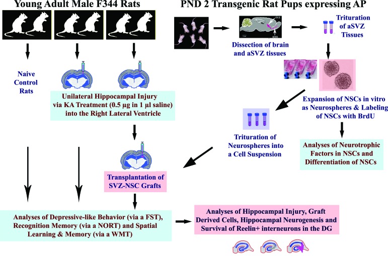
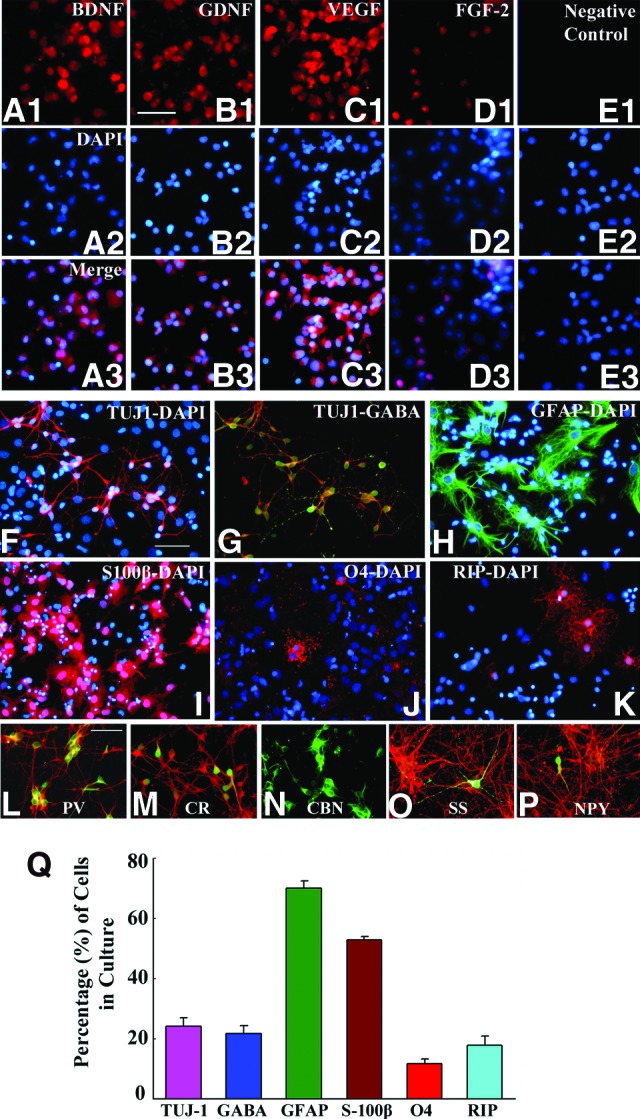
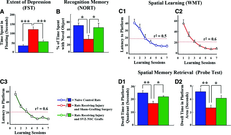
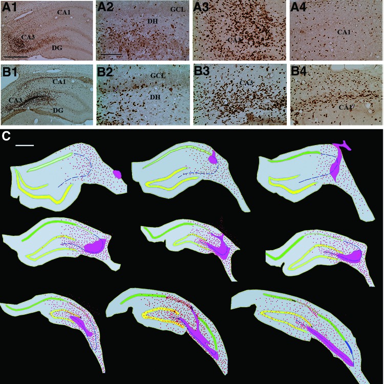
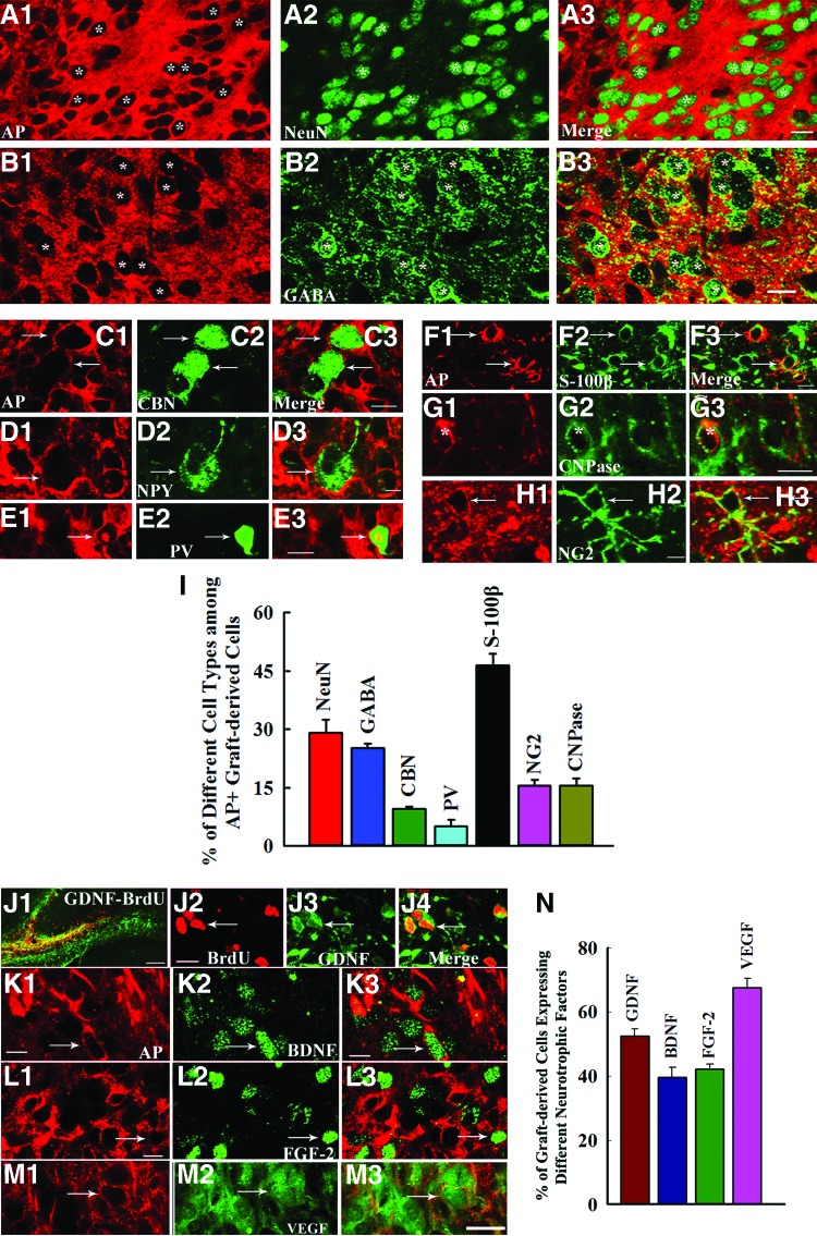
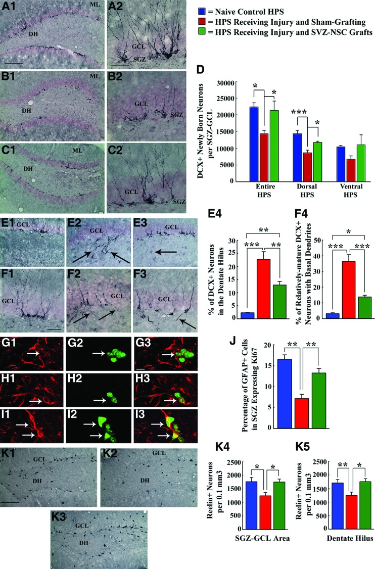
The hippocampus is vital for functions such as mood and memory. Hippocampal injury typically leads to mood and memory impairments associated with reduced and aberrant neurogenesis in the dentate gyrus. We examined whether neural stem cell (NSC) grafting after hippocampal injury would counteract impairments in mood, memory, and neurogenesis. We expanded NSCs from the anterior subventricular zone (SVZ) of postnatal F344 rat pups expressing the human placental alkaline phosphatase and grafted them into the hippocampus of young adult F344 rats at 5 days after an injury inflicted through a unilateral intracerebroventricular administration of kainic acid. Analyses through forced swim, water maze, and novel object recognition tests revealed significant impairments in mood and memory function in animals that underwent injury and sham-grafting surgery. In contrast, animals that received SVZ-NSC grafts after injury exhibited mood and memory function comparable to those of naïve control animals. Graft-derived cells exhibited excellent survival and pervasive migration, and they differentiated into neurons, subtypes of inhibitory GABAergic interneurons, astrocytes, oligodendrocytes, and oligodendrocyte progenitors. Significant fractions of graft-derived cells also expressed beneficial neurotrophic factors such as the glial cell line-derived neurotrophic factor, brain-derived neurotrophic factor, fibroblast growth factor, and vascular endothelial growth factor. Furthermore, SVZ-NSC grafting counteracted the injury-induced reductions and abnormalities in neurogenesis by both maintaining a normal level of NSC activity in the subgranular zone and providing protection to reelin+ interneurons in the dentate gyrus. These results underscore that early SVZ-NSC grafting intervention after hippocampal injury is efficacious for thwarting mood and memory dysfunction and abnormal neurogenesis.


Figures

Figure 1.
Figure 1.
An overview of the experimental design. The right side of the figure shows dissection of the anterior SVZ tissues from the PND 2 rat pups expressing the transgene alkaline phosphatase, trituration of SVZ tissues and expansion of NSCs as neurospheres, labeling of NSCs with BrdU, trituration of neurospheres into a cell suspension for grafting studies, and in vitro analyses. The left side of the figure depicts the induction of unilateral hippocampal injury and transplantation of SVZ-NSC grafts into the hippocampus; evaluation of animals for depressive-like behavior using an FST, recognition memory using a NORT, and spatial learning and memory using a WMT; and histological analyses of the host hippocampus for various analyses. Abbreviations: aSVZ, anterior subventricular zone; BrdU, 5′-bromodeoxyuridine; DG, dentate gyrus; FST, forced swim test; KA, kainic acid; NORT, novel object recognition test; NSC, neural stem cell; PND, postnatal day; SVZ, subventricular zone; WMT, water maze test.
Figure 2.
Figure 2.
Subventricular zone-neural stem cells (SVZ-NSCs) expanded in vitro demonstrate the expression of multiple neurotrophic factors and produce different central nervous system phenotypes, including the GABAergic interneurons. (A1–E3): NSCs showing the expression of BDNF (A1–A3), GDNF (B1–B3), VEGF (C1–C3), and FGF-2 (D1–D3). (A2–E2): NSC nuclei expressing DAPI. (E1–E3): Negative control samples of NSCs. Scale bar = 50 μm. (F–K): Differentiation of SVZ-NSC derived cells (following 7 days of incubation in a differentiation medium) into Tuj-1+ neurons (F), GABA+ neurons (G), GFAP+ astrocytes (H), S100β+ astrocytes (I), O4+ immature oligodendrocytes (J), and RIP+ mature oligodendrocytes (K). (L–P): Differentiation of SVZ-NSC cells in vitro into subtypes of GABAergic interneurons (shown in green) expressing parvalbumin (L), calretinin (M), calbindin (N), somatostatin (O), and neuropeptide Y (P). Scale bar = 50 μm. The bar chart in (Q) illustrates percentages of different types of neurons and glia derived from SVZ-NSCs following 7 days of incubation in a differentiation medium. Abbreviations: BDNF, brain-derived neurotrophic factor; CBN, calbindin; CR, calretinin; DAPI, 4′,6-diamidino-2-phenylindole; FGF-2, fibroblast growth factor-2; GDNF, glial cell line-derived neurotrophic factor; GFAP, glial fibrillary acidic protein; NPY, neuropeptide Y; PV, parvalbumin; RIP, receptor-interacting protein; SS, somatostatin; TUJ1, β-III-tubulin; VEGF, vascular endothelial growth factor.
Figure 3.
Figure 3.
Grafting of SVZ-NSCs into the hippocampus at 5 days postinjury maintains normal mood and memory function. (A): Results of an FST, which used the time spent in floating as a measure of depression. Rats receiving sham-grafting surgery after hippocampal injury (red) displayed increased depressive-like behavior, whereas rats receiving SVZ-NSC grafts after hippocampal injury (green) exhibited mood function similar to that of naïve control rats (blue). ***, p < .001. (B): Findings in a NORT, which used percentage of the exploration time spent with novel object as a measure of the object recognition memory. Rats receiving sham-grafting surgery exhibited impaired object recognition memory, whereas rats receiving SVZ-NSC grafts demonstrated object recognition ability comparable to that of naïve control rats. *, p < .05. (C1–D2): Results of a WMT. Note that based on changes in the mean latency values to reach the hidden platform over seven learning sessions (C1–C3), rats in all three groups exhibited ability for spatial learning (r2 = 0.5–0.6). (D1, D2): Comparison of memory retrieval function among the three groups, based on the amounts of time spent within the platform quadrant (D1) and the platform area (D2) in a probe test conducted at 24 hours after the seventh learning session. Note that rats receiving sham-grafting surgery after hippocampal injury exhibited impaired memory retrieval ability, whereas rats receiving SVZ-NSC grafts after hippocampal injury displayed memory retrieval ability similar to that of naïve control rats. *, p < .05; **, p < .01. Abbreviations: FST, forced swim test; NORT, novel object recognition test; SVZ-NSC, subventricular zone-neural stem cell; WMT, water maze test.
Figure 4.
Figure 4.
Cells derived from the subventricular zone-neural stem cell grafts migrate profusely into different regions of the injured hippocampus. (A1, A2): Examples of injured hippocampi that demonstrated pervasive migration of 5′-bromodeoxyuridine-labeled graft-derived cells. (A2–A4, B2-B4): Respectively, graft-derived cells in magnified regions of the dentate gyrus and the CA3 and CA1 subfields from (A1) and (A2). (C): Tracings of every 15th section through the hippocampus (performed using Neurolucida [MicroBrightField]) to show the distribution of graft-derived cells in one of the grafted animals. Note that the graft core regions (solid pink areas) are located in the lesioned CA3 subfield, whereas the graft-derived cells migrated into all three regions of the hippocampus. Scale bars = 500 μm (A1, B1, C), 200 μm (A2–A4, B2–B4). Abbreviations: CA, cornu ammonis; DG, dentate gyrus; DH, dentate hilus; GCL, granule cell layer.
Figure 5.
Figure 5.
Significant fractions of cells derived from the subventricular zone-neural stem cell (SVZ-NSC) grafts differentiate into different types of neurons and glia, and express several neurotrophic factors. (A1–H3): The AP-positive graft-derived cells (depicted in red) differentiated into neurons expressing NeuN (A1–A3); interneurons expressing GABA (B1–B3), calbindin (C1–C3), NPY (D1–D3), or PV (E1–E3); S100β+ astrocytes (F1–F3); CNPase+ oligodendrocytes (G1–G3); and NG2+ oligodendrocyte progenitors (H1–H3). The bar chart in (I) illustrates the percentages of different types of neurons and glia derived from SVZ-NSC grafts. (J1–M3): Cells derived from (BrdU+/AP+) SVZ-NSC grafts expressed GDNF (J1–J4), BDNF (K1–K3), FGF-2 (L1–L3), and VEGF (M1–M3). The bar chart in (N) illustrates the percentages of graft-derived cells expressing GDNF, BDNF, FGF-2, and VEGF. Scale bars = 10 μm (A1–H3, J2–M3), 100 μm (J1). Abbreviations: AP, alkaline phosphatase; BDNF, brain-derived neurotrophic factor; BrdU, 5′-bromodeoxyuridine; CBN, calbindin; CNPase, 2′,3′-cyclic nucleotide 3′-phosphodiesterase; FGF-2, fibroblast growth factor-2; GABA, γ-aminobutyric acid; GDNF, glial cell line-derived neurotrophic factor; NeuN, neuron-specific nuclear antigen; NG2, neuron glia proteoglycan 2; NPY, neuropeptide Y; PV, parvalbumin; VEGF, vascular endothelial growth factor.
Figure 6.
Figure 6.
Grafting of SVZ-NSCs into the HPS maintains neurogenesis and neural stem cell (NSC) activity at levels comparable to those of the intact control HPS and preserves reelin+ interneurons in the dentate gyrus. *, p < .05; **, p < .01; ***, p < .001. (A1–D): Extent of neurogenesis measured through DCX immunostaining. (E1–F4): Pattern of neurogenesis. (G1–J): NSC activity in the SGZ examined through GFAP-Ki67 dual immunofluorescence. (K1–K5): Reelin+ interneurons in the SGZ-GCL and the DH examined through reelin immunostaining. Note that in comparison with naïve control rats (A1, A2, D; E1, E4; F1, F4; G1–G3, J; K1, K4, K5), rats receiving sham-grafting surgery after hippocampal injury exhibited (a) decreased neurogenesis (B1, B2, D); (b) greater fractions of newly born neurons migrating abnormally into the dentate hilus (E2, E4); (c) increased occurrences of aberrant basal dendrites from newly born neurons (F2, F4); (d) decreased NSC activity (H1–H3, J); and (e) decreased numbers of reelin+ interneurons (K2, K4, K5). However, in, rats receiving SVZ-NSC grafts after hippocampal injury, the extent of neurogenesis (C1, C2, D), NSC activity (I1–I3, J), and surviving reelin+ interneuron numbers (K3, K4, K5) were comparable to those observed in naïve control rats. Additionally, both abnormal hilar migration of newly born neurons (E3, E4) and occurrences of aberrant basal dendrites (F3, F4) were greatly reduced in these rats. Scale bars = 200 μm (A1, B1, C1), 50 μm (A2, B2, C2, F1–F3), 100 μm (E1–E3), 10 μm (G1–I3), 200 μm (K1–K3). Abbreviations: DCX, doublecortin; DH, dentate hilus; GCL, granule cell layer; GFAP, glial fibrillary acidic protein; HPS, hippocampus; ML, molecular layer; SGZ, subgranular zone; SVZ-NSC, subventricular zone-neural stem cell.

Similar articles

Cited by

References

    1. Kryukov VI. The role of the hippocampus in long-term memory: Is it memory store or comparator? J Integr Neurosci. 2008;7:117–184. - PubMed
    1. Deng W, Aimone JB, Gage FH. New neurons and new memories: How does adult hippocampal neurogenesis affect learning and memory? Nat Rev Neurosci. 2010;11:339–350. - PMC - PubMed
    1. Samuels BA, Hen R. Neurogenesis and affective disorders. Eur J Neurosci. 2011;33:1152–1159. - PubMed
    1. Cohen AS, Pfister BJ, Schwarzbach E, et al. Injury-induced alterations in CNS electrophysiology. Prog Brain Res. 2007;161:143–169. - PubMed
    1. Huh JW, Widing AG, Raghupathi R. Differential effects of injury severity on cognition and cellular pathology after contusive brain trauma in the immature rat. J Neurotrauma. 2011;28:245–257. - PMC - PubMed

Publication types