. 2009 Oct;4(4):238-52.

Ocular Drug Delivery; Impact of in vitro Cell Culture Models

Affiliations

Ocular Drug Delivery; Impact of in vitro Cell Culture Models

Jaleh Barar et al. J Ophthalmic Vis Res. 2009 Oct.

Abstract

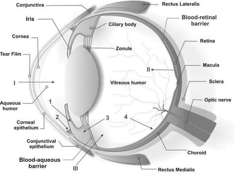
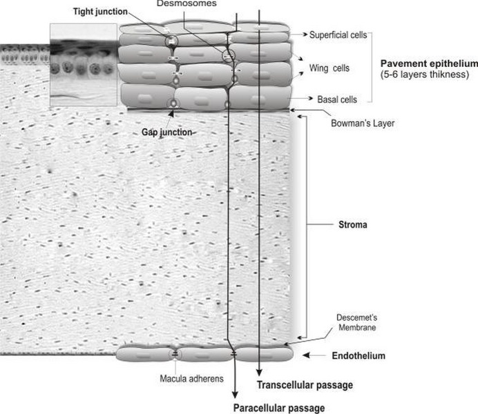
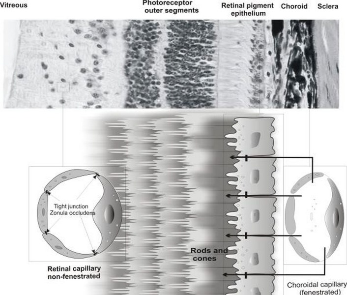
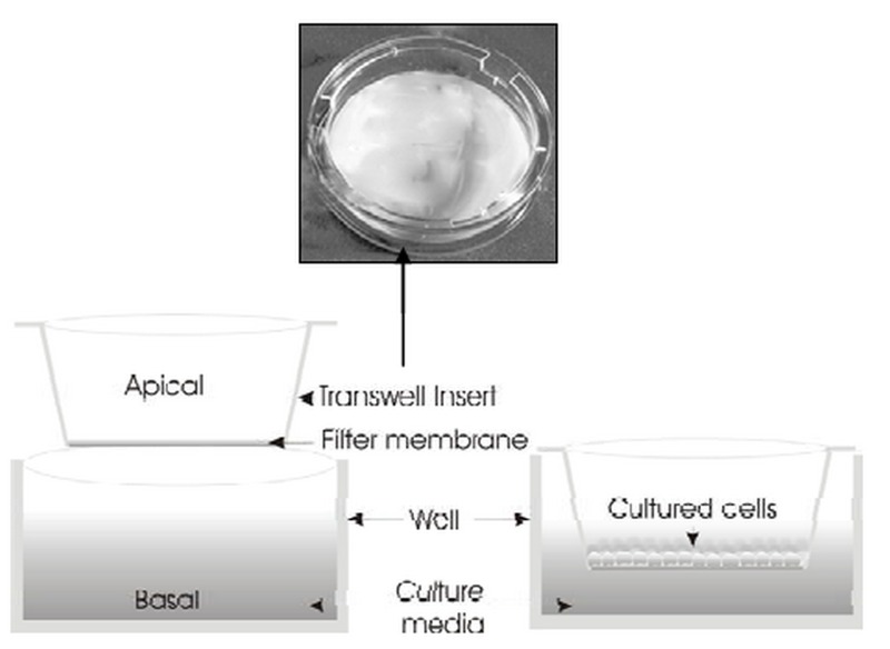
Normal vision depends on the optimal function of ocular barriers and intact membranes that selectively regulate the environment of ocular tissues. Novel pharmacotherapeutic modalities have aimed to overcome such biological barriers which impede efficient ocular drug delivery. To determine the impact of ocular barriers on research related to ophthalmic drug delivery and targeting, herein we provide a review of the literature on isolated primary or immortalized cell culture models which can be used for evaluation of ocular barriers. In vitro cell cultures are valuable tools which serve investigations on ocular barriers such as corneal and conjunctival epithelium, retinal pigment epithelium and retinal capillary endothelium, and can provide platforms for further investigations. Ocular barrier-based cell culture systems can be simply set up and used for drug delivery and targeting purposes as well as for pathological and toxicological research.

Keywords: Drug Delivery Systems; In Vitro.


Figures

Figure 1
Figure 1
Schematic illustration of ocular structures and barriers. The primary physiologic obstacle against topically instilled drugs is the tear film. The cornea is the main route for drug transport into the anterior chamber (I). The retinal pigment epithelium and the retinal capillary endothelium are main barriers against systemically administered drugs (II). Intravitreal injection is an invasive strategy to reach the vitreous (III). Administered drugs can be carried out of the anterior chamber by venous blood flow after diffusion across the iris surface (1) or by aqueous humor outflow (2). Drugs may be removed from the vitreous cavity through diffusion into the anterior chamber (3), or by the blood-retinal barrier (4).
Figure 2
Figure 2
Corneal cellular organization, the cornea consists of various transport limiting layers. The tightest monolayer is made by outer superficial epithelial cells which display tight junction complexes. The wing and basal cells exhibit gap junctions. The stroma and Descemet’s membrane cover the inner endothelial cells which contain macula adherens and are more permeable.
Figure 3
Figure 3
Retinal cellular architecture, retinal pigment epithelial (RPE) cells and retinal capillary endothelial (RCE) cells represent the outer and inner retinal barriers, respectively. RPE and RCE compose the main organization of the transport limiting layers. The outer layer of the retinal pigment epithelium displays tight barriers due to the presence of tight junctions (zonula occludens). Inner retinal capillary endothelial cells possess tight junctions and are non-fenestrated as opposed to choroidal capillary endothelial cells.
Figure 4
Figure 4
Schematic representation of the in vitro cell culture model. Transwell® insert filters are widely used for assessment of bioelectrical properties of target cells and screening for drug permeability and targeting.
Figure 5
Figure 5
Light microscopic images of brain capillary endothelial cells. A) Porcine brain capillary endothelial cells. B) Side-by-side coculture of ECV304 and C6 cells on top and underneath the Transwell® insert filter membrane.

References

    1. Barar J, Javadzadeh AR, Omidi Y. Ocular novel drug delivery: impacts of membranes and barriers. Expert Opin Drug Deliv. 2008;5:567–581. - PubMed
    1. Urtti A. Challenges and obstacles of ocular pharmacokinetics and drug delivery. Adv Drug Deliv Rev. 2006;58:1131–1135. - PubMed
    1. Hornof M, Toropainen E, Urtti A. Cell culture models of the ocular barriers. Eur J Pharm Biopharm. 2005;60:207–225. - PubMed
    1. Dartt DA, Hodges RR, Zoukhri D. Tear and Their Secretion. In: Fischbarg J, editor. The biology of the eye. New York: Academic Press; 2006. pp. 21–82.
    1. Fullard RJ, Tucker D. Tear protein composition and the effects of stimulus. Adv Exp Med Biol. 1994;350:309–314. - PubMed

LinkOut - more resources