

doi: 10.1155/2012/574125.
Epub 2012 Nov 11.
Familial florid cemento-osseous dysplasia: a rare manifestation in an Indian family
Affiliations
- PMID: 23198165
- PMCID: PMC3502783
- DOI: 10.1155/2012/574125
Item in Clipboard
Familial florid cemento-osseous dysplasia: a rare manifestation in an Indian family
Case Rep Dent.
2012.
Abstract
Florid cemento-osseous dysplasia (FCOD) is one of the uncommon dysplasias affecting the maxillofacial region. The age group may vary from 19 to 76 years and typically presents in the 4th and 5th decades. In most cases patients do not have hereditary basis of disease, and only a few familial cases have been documented. As far as we know this is the 1st reported case of familial FCOD in an Indian family. The mother and son exhibited multiple sclerotic masses in both jaws. The mode of transmission appeared to be autosomal dominant with variable phenotypic expression.
Figures

Extraoral photograph of patient (Case 1).
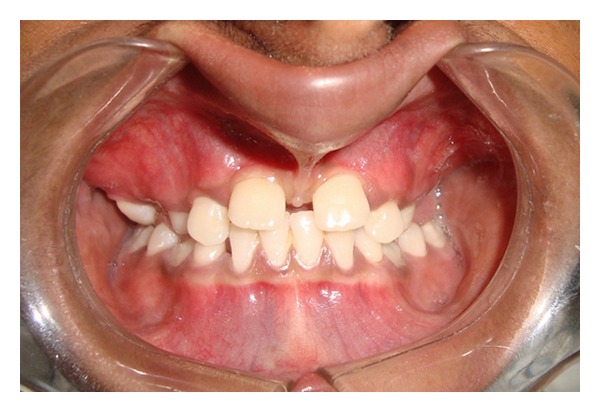
Intraoral photograph showing high frenal attachment (Case 1).

Intraoral photograph showing maxillary arch with retained deciduous teeth (Case 1).

Intraoral photograph showing mandibular arch (Case 1).

Panoramic radiograph (Case 1).

CT scan showing sections of lesion in the periapical region (Case 1).

CT scan showing panoramic like reconstruction (Case 1).

CT scan showing 3D reconstruction (Case 1).
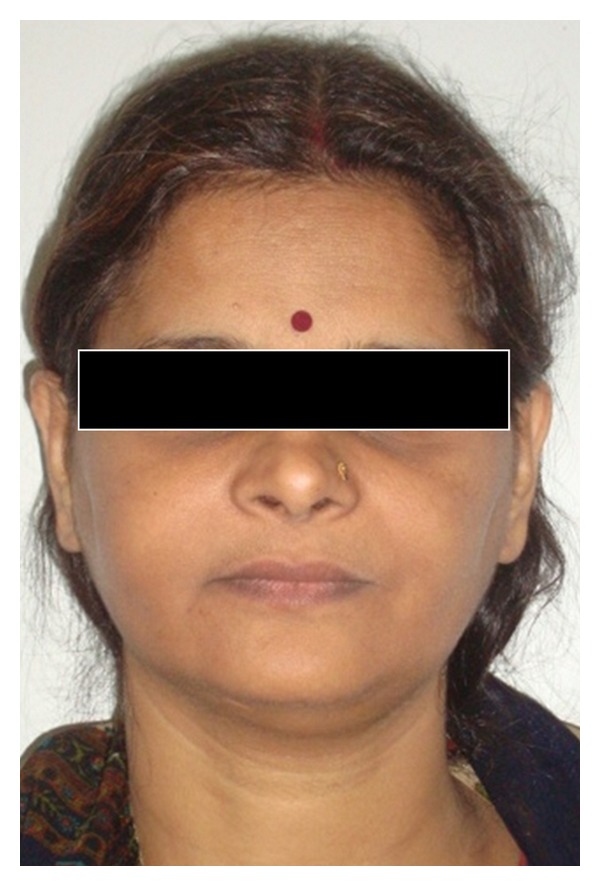
Extraoral photograph of patient's mother (Case 2).

Intraoral photograph of patient's mother (Case 2).

Intraoral photograph showing mandibular arch (Case 2).

Panoramic radiograph (Case 2).

CT scan of patient's mother (Case 2).

Photomicrograph (10x, H and E stained section) showing features of cemento-ossifying fibroma (Case 1).
References
-
- Said-al-Naief NA, Surwillo E. Florid osseous dysplasia of the mandible: report of a case. Compendium of Continuing Education in Dentistry. 1999;20(11):1017–1032. - PubMed
-
- Melrose RJ, Abrams AM, Mills BG. Florid osseous dysplasia. A clinical pathologic study of thirty four cases. Oral Surgery Oral Medicine and Oral Pathology. 1976;41(1):62–82. - PubMed
-
- Waldron CA. Fibro-osseous lesions of the jaws. Journal of Oral and Maxillofacial Surgery. 1985;43(4):249–262. - PubMed
-
- Jerjes W, Banu B, Swinson B, Hopper C. Florid cemento-osseous dysplasia in a young Indian woman. A case report. British Dental Journal. 2005;198(8):477–478. - PubMed
-
- Mangala M, Ramesh D, Surekha P, Santosh P. Florid cemento-osseous dysplasia: review and report of two cases. Indian Journal of Dental Research. 2006;17(3):131–134. - PubMed
LinkOut - more resources
Full Text Sources
Research Materials
