

A case report of a malignant peripheral nerve sheath tumor of the oral cavity in neurofibromatosis type 1
- PMID: 23198228
- PMCID: PMC3502806
- DOI: 10.1155/2012/936735
A case report of a malignant peripheral nerve sheath tumor of the oral cavity in neurofibromatosis type 1
Abstract
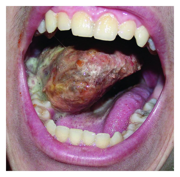
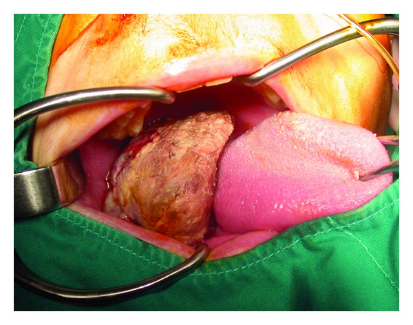
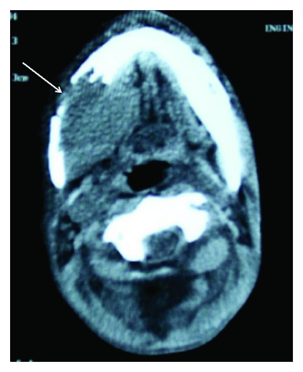
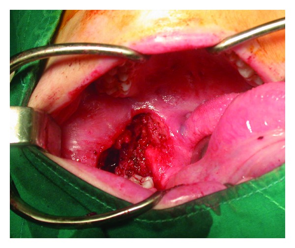
Patients with neurofibromatosis type 1 develop both benign and malignant tumors at an increased frequency. Most of the malignant peripheral nerve sheath tumors (MPNSTs) are considered as high-grade sarcomas originating from tissues of mesenchymal origin. It is generally accepted that MPNSTs occur in about 2% to 5% of neurofibromatosis patients. In this paper, we present a 16-year-old male patient with neurofibromatosis who developed MPNST of the retromolar area. The mass enlarged rapidly in a period of 6 weeks. The patient was treated surgically, and a tumor mass with a diameter of 7 × 6 × 4 cm was excised, but after 8 months a recurrence was observed at the same site. The sarcomatous change in a neurofibroma has an extremely poor prognosis, so patients with neurofibromatosis should be closely monitored for a possible malignancy. A rapid change in size of a preexisting neurofibroma, infiltration of the adjacent structures, intralesional hemorrhage, and pain indicate a possible malignant transformation to MPNST.
Figures
References
-
- Neurofibromatosis. Conference statement. National Institutes of Health Consensus Development Conference. Archives of Neurology. 1988;45:575–578. - PubMed
-
- Ferner RE, Gutmann DH. International consensus statement on malignant peripheral nerve sheath tumors in neurofibromatosis. Cancer Research. 2002;62(5):1573–1577. - PubMed
-
- Munhoz EA, Cardoso CL, Tolentino ES, et al. Von Recklinghausen's disease- diagnosis from oral lesion. neurofibromatosis 1. International Journal of Odontostomatology. 2010;4:179–183.
-
- D’Ambrosio JA, Langlais RP, Young RS. Jaw and skull changes in neurofibromatosis. Oral Surgery Oral Medicine and Oral Pathology. 1988;66(3):391–396. - PubMed
LinkOut - more resources
Full Text Sources
Research Materials
