. 2013 Feb;118(2):465-77.
doi: 10.3171/2012.10.JNS111836. Epub 2012 Nov 30.

Evaluation of the hematoma consequences, neurobehavioral profiles, and histopathology in a rat model of pontine hemorrhage

Affiliations

Evaluation of the hematoma consequences, neurobehavioral profiles, and histopathology in a rat model of pontine hemorrhage

Tim Lekic et al. J Neurosurg. 2013 Feb.

Abstract

Object: Primary pontine hemorrhage (PPH) represents approximately 7% of all intracerebral hemorrhages (ICHs) and is a clinical condition of which little is known. The aim of this study was to characterize the early brain injury, neurobehavioral outcome, and long-term histopathology in a novel preclinical rat model of PPH.

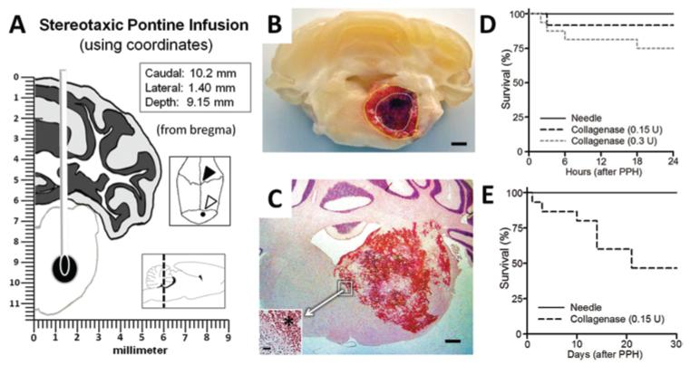
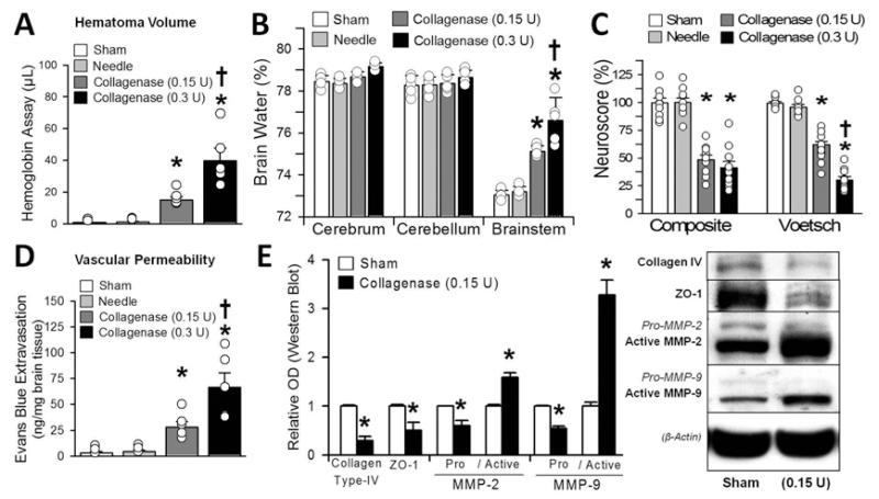
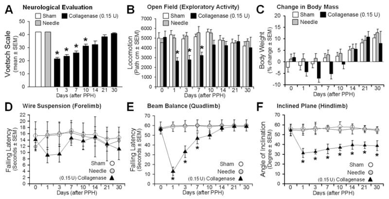
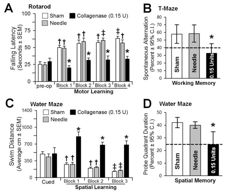
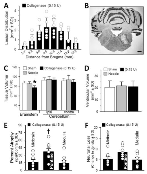
Methods: The authors stereotactically infused collagenase (Type VII) into the ventral pontine tegmentum of the rats, in accordance with the most commonly affected clinical region. Measures of cerebrovascular permeability (brain water content, hemoglobin assay, Evans blue, collagen Type IV, ZO-1, and MMP-2 and MMP-9) and neurological deficit were quantified at 24 hours postinfusion (Experiment 1). Functional outcome was measured over a 30-day period using a vertebrobasilar scale (the modified Voetsch score), open field, wire suspension, beam balance, and inclined-plane tests (Experiment 2). Neurocognitive ability was determined at Week 3 using the rotarod (motor learning), T-maze (working memory), and water maze (spatial learning and memory) (Experiment 3), followed by histopathological analysis 1 week later (Experiment 4).

Results: Stereotactic collagenase infusion caused dose-dependent elevations in hematoma volume, brain edema, neurological deficit, and blood-brain barrier rupture, while physiological variables remained stable. Functional outcomes mostly normalized by Week 3, whereas neurocognitive deficits paralleled the cystic cavitary lesion at 30 days. Obstructive hydrocephalus did not develop despite a clinically relevant 30-day mortality rate (approximately 54%).

Conclusions: These results suggest that the model can mimic several translational aspects of pontine hemorrhage in humans and can be used in the evaluation of potential preclinical therapeutic interventions.


Figures

Fig. 1
Fig. 1
Experimental model of pontine hemorrhage in the rat. A: Schematic showing stereotactic needle placement into the right ventral pontine tegmentum (Insets: coordinates [upper]; cranial aspect of bregma [open arrowhead] and the infusion site [closed arrowhead]; sagittal demonstration with the dotted line at craniocaudal level of infusion [lower]). B: Photograph of coronal section of tissue at 24 hours after collagenase (0.15 U) infusion, showing the hematoma surrounded by vasogenic edema (outlined with dotted lines). Bar = 1 mm. C: Representative H & E-stained cryosection (12 hours, 0.3 U collagenase) illustrating the hemorrhage. Bar = 0.5 mm. (Inset showing the hematoma border [asterisk]. Bar = 20 μm.) D and E: Gehan-Breslow survival analysis results at 24 hours and 30 days.
Fig. 2
Fig. 2
Hematoma consequences at 24 hours: hematoma volume assay (A), brain water (B), neuroscore (composite and Voetsch) (C), vascular permeability (D), and (E) semiquantification (left) of the collagen Type IV, ZO-1, and MMP-2 and -9 protein immunoblots (right). Values are expressed as the mean ± SEM (n = 10 [neuroscore] and n = 5 [all others]). *p < 0.05 compared with controls (sham and needle trauma). †p < 0.05 compared with collagenase infusion (0.15 U). Circles in bars indicate the raw data points of each measurement, and whiskers represent the SEM. OD = optical density.
Fig. 3
Fig. 3
Neurofunctional profiles in 30-day period: Voetsch score (A), locomotion (B), body weight (C), wire suspension (D), beam balance (E), and inclined plane (F). Values are expressed as mean ± SEM (n = 8 [sham and needle trauma] and n = 7 [collagenase infusion, 0.15 units]). *p < 0.05 comparing collagenase infusion (closed triangles) with controls (sham, or needle trauma, open circles). path = path length.
Fig. 4
Fig. 4
Neurocognitive ability at 3 weeks. A: Motor learning assessed by the change in the latency to falling off an accelerating rotarod (2 rpm/5 sec) preoperatively and across 4 daily blocks. B: Working memory quantified by the number of spontaneous alternations in the T-maze. C: Spatial learning assessed by the swim distance needed to find the visible (cued) versus the hidden (spatial) platforms in the water maze. D: Spatial memory was determined by the percent duration in the probe quadrant when the platform was removed. Values expressed as the mean ± SEM (rotarod, cued and spatial water maze) and mean ± 95% CI (T-maze and probe quadrant), n = 8 (sham and needle trauma), n = 7 (collagenase infusion, 0.15 U). *p < 0.05 compared with controls (sham and needle trauma). †p < 0.05 compared with preoperative (rotarod) or cued trials (spatial water maze). ‡ p < 0.05 compared with Block 1 (rotarod and spatial water maze).
Fig. 5
Fig. 5
Histopathological results at 30 days. A: Lesion distribution (in mm2) throughout the brainstem. B: Representative cryosection illustrating the cystic cavitary lesion. Bar = 1 mm. C: Infratentorial brain tissue volume (in mm3). D: Volume of the ventricles (in mm3). E: Percentage of atrophy of the brainstem (midbrain, pontine, and medulla), expressed as the volumetric difference between ipsilateral and contralateral sides. F: Neuronal density loss over the intact (perilesional) brainstem regions expressed as average difference in cell counts between ipsilateral and contralateral sides. Values expressed as the mean ± SD (n = 8 [sham and needle trauma] and n = 7 [collagenase infusion, 0.15 U]). *p < 0.05 compared with controls (sham and needle). †p < 0.05 compared with midbrain and medulla (percentage of atrophy).

Comment in

  • Letter to the Editor: pontine hemorrhage.
    Tsai YC, Liu FC, Chang CM, Chang CH, Liao YH, Yang TB, Hueng DY. Tsai YC, et al. J Neurosurg. 2013 May;118(5):1152. doi: 10.3171/2012.12.JNS122420. Epub 2013 Mar 22. J Neurosurg. 2013. PMID: 23521553 No abstract available.
  • Response.
    Lekic T, Rolland W, Krafft PR, Suzuki H, Tang J, Zhang JH, Kamper JE, Hartman RE. Lekic T, et al. J Neurosurg. 2013 May;118(5):1152-3. J Neurosurg. 2013. PMID: 23776937 No abstract available.

References

    1. Andaluz N, Zuccarello M, Wagner KR. Experimental animal models of intracerebral hemorrhage. Neurosurg Clin N Am. 2002;13:385–393. - PubMed
    1. Avendaño C, Machín R, Bermejo PE, Lagares A. Neuron numbers in the sensory trigeminal nuclei of the rat: A GABA- and glycine-immunocytochemical and stereological analysis. J Comp Neurol. 2005;493:538–553. - PubMed
    1. Balci K, Asil T, Kerimoglu M, Celik Y, Utku U. Clinical and neuroradiological predictors of mortality in patients with primary pontine hemorrhage. Clin Neurol Neurosurg. 2005;108:36–39. - PubMed
    1. Batton RR, III, Jayaraman A, Ruggiero D, Carpenter MB. Fastigial efferent projections in the monkey: an autoradiographic study. J Comp Neurol. 1977;174:281–305. - PubMed
    1. Bermejo PE, Jiménez CE, Torres CV, Avendaño C. Quantitative stereological evaluation of the gracile and cuneate nuclei and their projection neurons in the rat. J Comp Neurol. 2003;463:419–433. - PubMed

Publication types

LinkOut - more resources