

The GGGGCC repeat expansion in C9ORF72 in a case with discordant clinical and FDG-PET findings: PET trumps syndrome
- PMID: 23199140
- PMCID: PMC3593970
- DOI: 10.1080/13554794.2012.732090
The GGGGCC repeat expansion in C9ORF72 in a case with discordant clinical and FDG-PET findings: PET trumps syndrome
Abstract
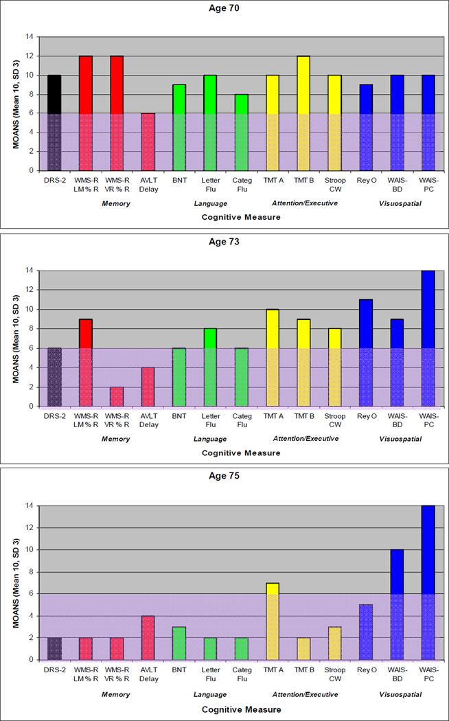
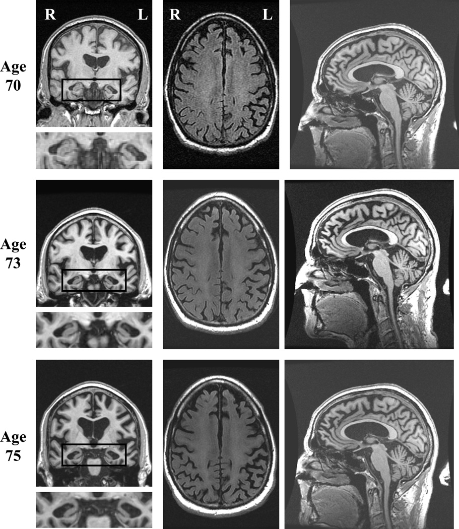
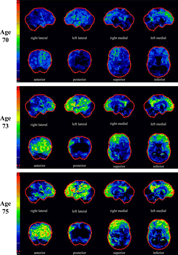
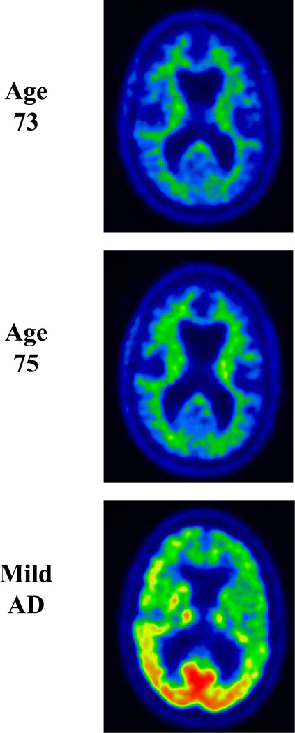
A hexanucleotide repeat expansion in the chromosome 9 open reading frame 72 (C9ORF72) gene was recently discovered as the cause underlying frontotemporal degeneration (FTD) and/or amyotrophic lateral sclerosis (ALS) linked to chromosome 9 (c9FTD/ALS). In this atypical case of c9FTD/ALS, the proband presented with amnestic mild cognitive impairment which evolved into Alzheimer's disease (AD)-type dementia and later developed ALS. Fluorodeoxyglucose-positron emission tomography of the brain demonstrated mild hypometabolism involving the medial frontal and lateral temporal lobes, left more so than right, which progressed over time. He was subsequently confirmed to have the C9ORF72 expansion. This report highlights the need to consider mutations in the FTD-associated genes when a familial disorder is suggested and neuroimaging studies reveal findings atypical of an AD pathophysiological process despite the typical anterograde amnestic syndrome.
Figures
Similar articles
-
Brain ¹⁸F-FDG and ¹¹C-PiB PET findings in two siblings with FTD/ALS associated with the C9ORF72 repeat expansion.Neurocase. 2014 Apr;20(2):150-7. doi: 10.1080/13554794.2012.741252. Epub 2012 Dec 5. Neurocase. 2014. PMID: 23216213
-
Characterization of a family with c9FTD/ALS associated with the GGGGCC repeat expansion in C9ORF72.Arch Neurol. 2012 Sep;69(9):1164-9. doi: 10.1001/archneurol.2012.772. Arch Neurol. 2012. PMID: 22637471 Free PMC article.
-
The metabolic signature of C9ORF72-related ALS: FDG PET comparison with nonmutated patients.Eur J Nucl Med Mol Imaging. 2014 May;41(5):844-52. doi: 10.1007/s00259-013-2667-5. Epub 2014 Jan 21. Eur J Nucl Med Mol Imaging. 2014. PMID: 24445987 Free PMC article.
-
Molecular Mechanisms of Neurodegeneration Related to C9orf72 Hexanucleotide Repeat Expansion.Behav Neurol. 2019 Jan 15;2019:2909168. doi: 10.1155/2019/2909168. eCollection 2019. Behav Neurol. 2019. PMID: 30774737 Free PMC article. Review.
-
Insights into the pathogenic mechanisms of Chromosome 9 open reading frame 72 (C9orf72) repeat expansions.J Neurochem. 2016 Aug;138 Suppl 1:145-62. doi: 10.1111/jnc.13623. Epub 2016 Jun 15. J Neurochem. 2016. PMID: 27016280 Review.
Cited by
-
Biomarkers for Amyotrophic Lateral Sclerosis and Frontotemporal Dementia Associated With Hexanucleotide Expansion Mutations in C9orf72.Front Neurol. 2018 Dec 5;9:1063. doi: 10.3389/fneur.2018.01063. eCollection 2018. Front Neurol. 2018. PMID: 30568632 Free PMC article. Review.
-
Phenotypic variability and neuropsychological findings associated with C9orf72 repeat expansions in a Bulgarian dementia cohort.PLoS One. 2018 Dec 14;13(12):e0208383. doi: 10.1371/journal.pone.0208383. eCollection 2018. PLoS One. 2018. PMID: 30550541 Free PMC article.
-
Heterogeneous brain FDG-PET metabolic patterns in patients with C9orf72 mutation.Neurol Sci. 2019 Mar;40(3):515-521. doi: 10.1007/s10072-018-3685-7. Epub 2018 Dec 15. Neurol Sci. 2019. PMID: 30554355
-
C9ORF72 mutations in neurodegenerative diseases.Mol Neurobiol. 2014 Feb;49(1):386-98. doi: 10.1007/s12035-013-8528-1. Epub 2013 Aug 10. Mol Neurobiol. 2014. PMID: 23934648 Review.
-
Phenotypic Heterogeneity of Monogenic Frontotemporal Dementia.Front Aging Neurosci. 2015 Sep 1;7:171. doi: 10.3389/fnagi.2015.00171. eCollection 2015. Front Aging Neurosci. 2015. PMID: 26388768 Free PMC article. Review.
References
-
- Arai T, Hasegawa M, Akiyama H, Ikeda K, Nonaka T, Mori H, et al. TDP-43 is a component of ubiquitin-positive tau-negative inclusions in frontotemporal lobar degeneration and amyotrophic lateral sclerosis. Biochem Biophys Res Comm. 2006;351:602–611. - PubMed
-
- Brooks BR, Miller RG, Swash M, Munsat TL. El Escorial revisited: revised criteria for the diagnosis of amyotrophic lateral sclerosis. Amyotrophic Lateral Sclerosis and Other Motor Neuron Disorders. 2000;1:293–299. - PubMed
Publication types
MeSH terms
Substances
Grants and funding
LinkOut - more resources
Full Text Sources
Medical
Miscellaneous