. 2013 Jul;8(7):1055-65.
doi: 10.2217/nnm.12.138. Epub 2012 Dec 2.

Lysosome-targeted octadecyl-rhodamine B-liposomes enhance lysosomal accumulation of glucocerebrosidase in Gaucher's cells in vitro

Affiliations

Lysosome-targeted octadecyl-rhodamine B-liposomes enhance lysosomal accumulation of glucocerebrosidase in Gaucher's cells in vitro

Ritesh Thekkedath et al. Nanomedicine (Lond). 2013 Jul.

Abstract

Aim: We hypothesized that liposomes modified with lysosomotropic octadecyl-rhodamine B (Rh) and loaded with therapeutic glucocerebroside velaglucerase alfa (VPRIV™) will improve lysosomal delivery of the enzyme into Gaucher's cells.

Materials & methods: Confocal microscopy and flow cytometry were used to evaluate the ability of Rh-modified liposomes loaded with VPRIV to improve the lysosomal targeting in monocyte-derived macrophages and Gaucher's fibroblasts.

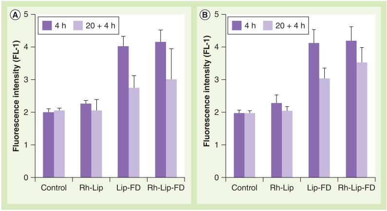
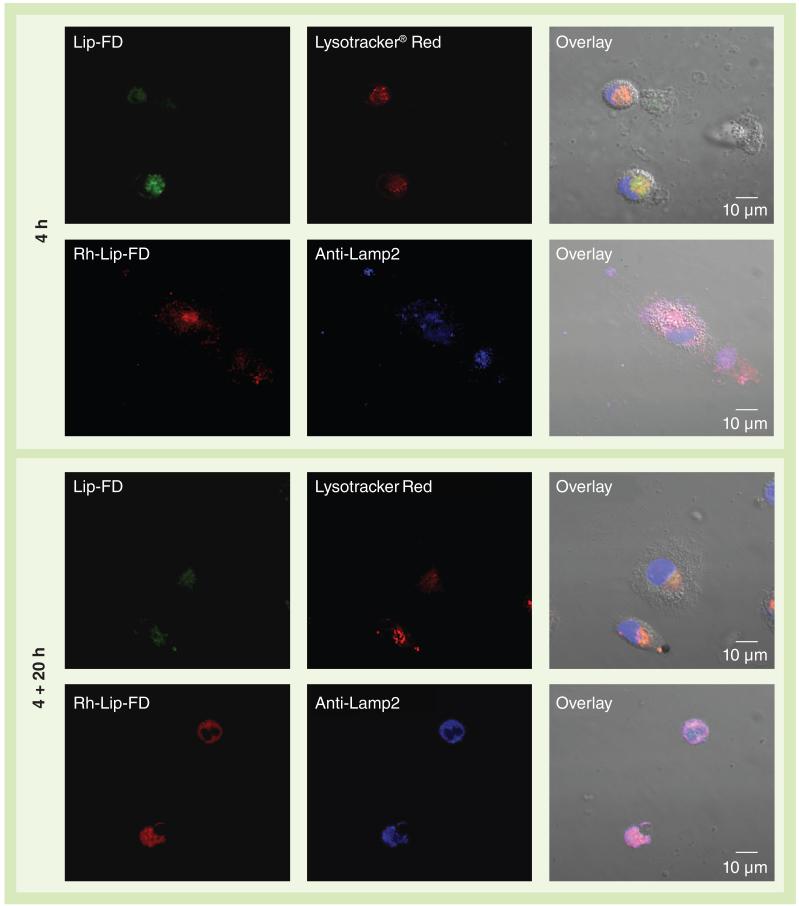
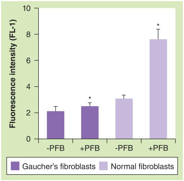
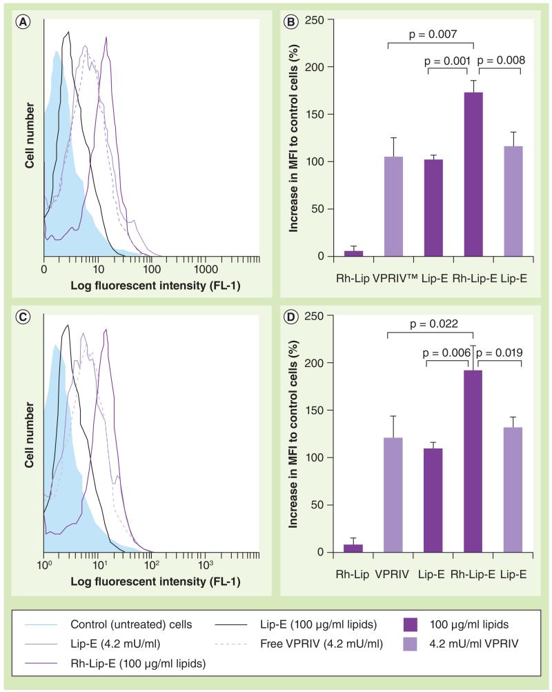
Results: Confocal microscopy demonstrated that Rh-modified liposomes localized primarily in the lysosomes. As confirmed by flow cytometry using specific substrate 5-(pentafluorobenzoylamino)fluorescein diglucoside, intralysosomal accumulation of VPRIV in the cells treated with Rh-modified liposomes was significantly increased (up to 68%) relative to the cells treated with plain liposomes or free VPRIV.

Conclusion: Rh-modified lysosomotropic liposomes can improve lysosomal accumulation of liposomal enzymes both in nonphagocytic Gaucher's fibroblasts and phagocytic monocyte-derived macrophages.


Figures

Figure 1
Figure 1. Post-conduritol B epoxide recovery of glucocerebrosidase activity in monocyte-derived macrophages
Monocyte-derived macrophages preincubated with conduritol B epoxide (CBE) for 72 h were post-incubated at 1, 24, 48 and 72 h with CBE-free medium. The recovery of the enzyme activity was measured by flow cytometry using 5-(pentafluorobenzoylamino)fluorescein diglucoside, a specific fluorogenic GlcCerase-specific substrate, and represented as a percentage of the enzyme activity in the control (CBE-untreated) cells. The data represent the mean of three different experiments ± standard deviation. GlcCerase: Glucocerebrosidase.
Figure 2
Figure 2. Interaction of liposomes with monocyte-derived macrophages
Macrophages had (A) active or (B) inhibited lysosomal glucocerebrosidase. Activity of glucocerebrosidase in monocyte-derived macrophages was inhibited by treatment of the cells with conduritol B epoxide (200 μM). The cells were treated with equal amounts of plain Lip-FD, Rh-Lip or Rh-Lip-FD for 4 h (dark bars) followed by washing and additional incubation for 20 h in liposome-free medium (pale bars). The cells were then treated with 5-(pentafluorobenzoylamino)fluorescein diglucoside and analyzed by flow cytometry. The data represent the mean of three different experiments ± standard deviation. Lip-FD: Fluorescein isothiocyanate-dextran-loaded plain liposomes; Rh-Lip: Octadecyl-rhodamine B-modified liposomes; Rh-Lip-FD: Fluorescein isothiocyanate-dextran-loaded octadecyl-rhodamine B-modified liposomes.
Figure 3
Figure 3. Co-localization of liposomal formulations with lysosomal markers
Monocyte-derived macrophages were treated with Lip-FD (green) or Rh-Lip-FD (red) for 4 h, followed by incubation for 20 h in the liposome-free medium. The treated cells were stained with lysosomal markers: Lysotracker® Red (Invitrogen/Molecular Probes Inc., OR, USA), shown here in red, or Lamp2 monoclonal antibodies (blue) in the case of Lip-FD-or Rh-Lip-FD-treated cells, respectively. In Lysotracker Red-stained cells, the nuclei were visualized by Hoechst 33342 (blue). Co-localization of liposomal formulations with respective lysosomal markers was analyzed by confocal microscopy. Scale bar = 10 μm. Lip-FD: Fluorescein isothiocyanate-dextran-loaded plain liposomes; Rh-Lip-FD: Fluorescein isothiocyanate-dextran-loaded octadecyl-rhodamine B-modified liposomes.
Figure 4
Figure 4. Glucocerebrosidase activity in Gaucher’s fibroblasts by flow cytometry
Gaucher’s and normal human fibroblasts were treated with PFB diglucoside and resultant fluorescence intensity (channel FL-1) was determined by flow cytometry. Each value is the mean ± standard deviation of three different experiments. *p < 0.0001. PFB: 5-(Pentafluorobenzoylamino)fluorescein.
Figure 5
Figure 5. Lysosomal delivery of velaglucerase alfa by octadecyl-rhodamine B-modified liposomes
Monocyte-derived macrophages with inhibited glucocerebrosidase (A & B) or Gaucher’s fibroblasts (C & D) were treated with Rh-Lip, Lip-E or Rh-Lip-E, or with free VPRIV for 4 + 20 h. The level of the intralysosomal glucocerebrosidase was measured by flow cytometry (channel FL-1) of the cells incubated with 5-(pentafluorobenzoylamino)fluorescein diglucoside. (A & C) Typical histogram analysis. (B & D) A percentage increase in MFI normalized to control (untreated) cells. Each value is the mean ± standard deviation of three different experiments (each in triplicate). Lip-E: Velaglucerase alfa-loaded plain liposomes; MFI: Mean fluorescent intensity; Rh-Lip: Octadecyl-rhodamine B-modified liposomes; Rh-Lip-E: Velaglucerase alfa-loaded rhodamine B-modified liposomes; VPRIV: Velaglucerase alfa.

References

    1. Grabowski GA. Phenotype, diagnosis, and treatment of Gaucher’s disease. Lancet. 2008;372(9645):1263–1271. - PubMed
    1. Van Der Ploeg AT, Reuser AJ. Pompe’s disease. Lancet. 2008;372(9646):1342–1353. - PubMed
    1. Zarate YA, Hopkin RJ. Fabry’s disease. Lancet. 2008;372(9647):1427–1435. - PubMed
    1. Grabowski GA, Hopkin RJ. Enzyme therapy for lysosomal storage disease: principles, practice, and prospects. Ann. Rev. Genomics Hum. Genet. 2003;4:403–436. - PubMed
    1. Jmoudiak M, Futerman AH. Gaucher disease: pathological mechanisms and modern management. Br. J. Haematol. 2005;129(2):178–188. - PubMed

Publication types