. 2012 Dec 3;3(1):17.
doi: 10.1186/1878-5085-3-17.

Retinitis pigmentosa and ocular blood flow

Affiliations

Retinitis pigmentosa and ocular blood flow

Katarzyna Konieczka et al. EPMA J. .

Abstract

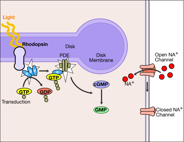
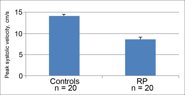
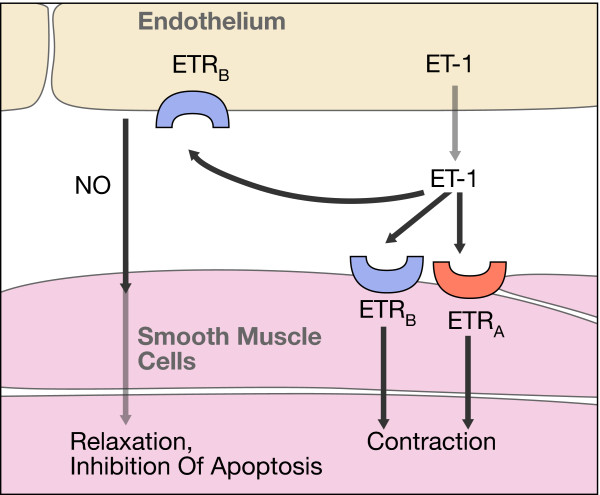
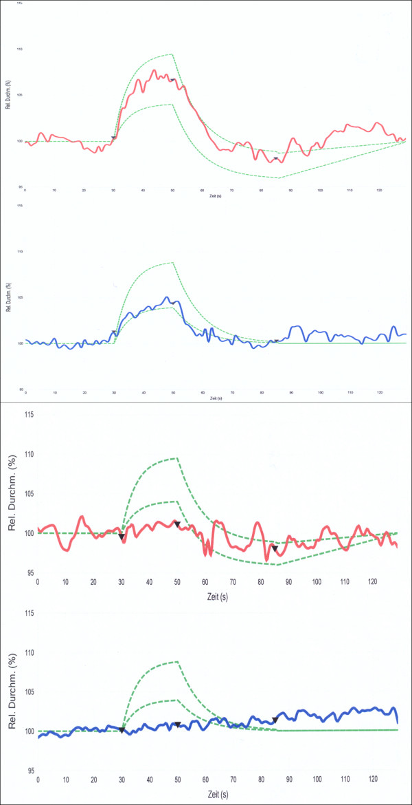
Is the concept of integrative, preventive and personalised medicine applicable to the relationship between retinitis pigmentosa (RP) and ocular blood flow (OBF)? RP encompasses a group of hereditary diseases of the posterior segment of the eye characterised by degeneration, atrophy and finally loss of photoreceptors and retinal pigment epithelium, leading to progressive visual loss. Many different mutations affecting different genes can lead to the clinical picture of RP. Even though the disease has a clear genetic background, there are obviously other factors influencing the manifestation and progression of RP. In this review, we focus on the role of OBF. There is evidence that, in PR patients, OBF is more reduced than one would expect secondary to the retinal atrophy. The main cause of this additional component seems to be primary vascular dysregulation (PVD) syndrome. As PVD syndrome is partly treatable, a vascular evaluation of RP patients is meaningful. Based on the outcome, a targeted individualised, preventive or supportive treatment might be introduced in selected RP patients.


Figures

Figure 1
Figure 1
Fundus photograph of a patient with retinitis pigmentosa with the typical bone-spicule pigmentary changes (arrows). Left: photograph; right: fluorescein angiogram.
Figure 2
Figure 2
Goldmann visual fields of a RP patient demonstrating progressive concentric constriction. Top: mid-stage RP; bottom: late-stage RP.
Figure 3
Figure 3
Octopus perimetry demonstrating the extensive visual field loss. Left: mid-stage RP; right: late-stage RP.
Figure 4
Figure 4
Fundus photograph in patient with late-stage RP with the classic triad. Bone-spicule retinal pigmentation (black arrow), retinal vessel attenuation (white arrow) and waxy disc pallor (red arrow).
Figure 5
Figure 5
Scotopic electroretinography (ERG), ISCEV standard (International Society for Clinical Electrophysiology of Vision). Normal ERG responses in healthy subject (left) and extinguished responses in RP patient (right).
Figure 6
Figure 6
Multifocal electroretinogram. Severely attenuated paracentral responses in RP patient (left) in comparison to normal responses in a healthy subject (right).
Figure 7
Figure 7
Dark adaptation. Dark adaptation of rods (green line) and cones (red line) in a healthy subject (top) and disturbed dark adaptation (loss of red-green dissociation) in RP patient (bottom).
Figure 8
Figure 8
Cystoid macular oedema in RP patient. Left: fundus photograph; right: optical coherence tomography image.
Figure 9
Figure 9
Phototransduction in rod photoreceptors. Mutations of genes involved in this process can lead to RP (Adapted from [14] with permission).
Figure 10
Figure 10
Histological section through the human retina. Left: healthy retina; middle: retina of a patient with mid-stage RP; right: retina of a patient with late-stage RP.
Figure 11
Figure 11
Peak systolic velocities (mean ± SEM) in posterior ciliary arteries. Control subjects (left) and patients with early-stage RP (right) (based on [2]).
Figure 12
Figure 12
Endothelin. ET-1 arises by cleaving the precursor molecules (Adapted from [14] with permission).
Figure 13
Figure 13
Endothelin receptors. In humans, the effects of ET-1 are mediated by two types of ET receptors: the type-A receptor (ETA) and the type-B receptor (ETB) (Adapted from [14] with permission).
Figure 14
Figure 14
Thermography of hands and faces. A subject without PVD (left) and a subject with PVD (right) (Adapted from [32] with permission).
Figure 15
Figure 15
The nailfold capillary microscopy examination. Left: examination setting with cooling device; middle: picture from a video with normal capillary blood flow; right: picture from the video demonstrating blood flow cessation after cold provocation in a PVD subject (Adapted from [32] with permission).
Figure 16
Figure 16
Outcome of blood pressure monitoring. Yellow: normal range; blue: systolic, diastolic and mean blood pressure of a RP patient.
Figure 17
Figure 17
Vascular response to light measured with a Retinal Vessel Analyser. Top: normal responses of both arteries and veins; bottom: reduced responses of both arteries and veins in a RP patient. Red lines: arteries; blue lines: veins.

References

    1. Ferrari S, Di Iorio E, Barbaro V, Ponzin D, Sorrentino FS, Parmeggiani F. Retinitis pigmentosa: genes and disease mechanisms. Curr Genomics. 2011;12:238–249. doi: 10.2174/138920211795860107. - DOI - PMC - PubMed
    1. Cellini M, Strobbe E, Gizzi C, Campos EC. ET-1 plasma levels and ocular blood flow in retinitis pigmentosa. Can J Physiol Pharmacol. 2010;88:630–635. doi: 10.1139/Y10-036. - DOI - PubMed
    1. Grunwald JE, Maguire AM, Dupont J. Retinal hemodynamics in retinitis pigmentosa. Am J Ophthalmol. 1996;122:502–508. - PubMed
    1. Falsini B, Anselmi GM, Marangoni D, D'Esposito F, Fadda A, Di Renzo A, Campos EC, Riva CE. Subfoveal choroidal blood flow and central retinal function in retinitis pigmentosa. Invest Ophthalmol Vis Sci. 2011;52:1064–1069. doi: 10.1167/iovs.10-5964. - DOI - PubMed
    1. Chizzolini M, Galan A, Milan E, Sebastiani A, Costagliola C, Parmeggiani F. Good epidemiologic practice in retinitis pigmentosa: from phenotyping to biobanking. Curr Genomics. 2011;12:260–266. doi: 10.2174/138920211795860071. - DOI - PMC - PubMed