

Review
doi: 10.1186/1479-7364-6-26.
The human crystallin gene families
Affiliations
- PMID: 23199295
- PMCID: PMC3554465
- DOI: 10.1186/1479-7364-6-26
Item in Clipboard
Review
The human crystallin gene families
Hum Genomics.
.
Abstract
Crystallins are the abundant, long-lived proteins of the eye lens. The major human crystallins belong to two different superfamilies: the small heat-shock proteins (α-crystallins) and the βγ-crystallins. During evolution, other proteins have sometimes been recruited as crystallins to modify the properties of the lens. In the developing human lens, the enzyme betaine-homocysteine methyltransferase serves such a role. Evolutionary modification has also resulted in loss of expression of some human crystallin genes or of specific splice forms. Crystallin organization is essential for lens transparency and mutations; even minor changes to surface residues can cause cataract and loss of vision.
Figures

Exon/intron structure of mammalian genes for α-, β- and γ-crystallins. Exons are shown as boxes with ORF in thicker boxes. CRYB genes coding for β-crystallins vary in 5′-exon structure. The exons encoding the four structural motifs of the β-crystallin and γ-crystallin proteins are indicated by 1, 2, 3, and 4. Some mammals have an alternatively spliced exon in the first intron of CRYAA, but in humans this is a pseudoexon.

Phylogenetic tree of the human sHSP gene family. Sequences were extracted from the UCSC web browser. Translated ORFs were aligned and neighbor-joining trees were constructed using MEGA4 [15].
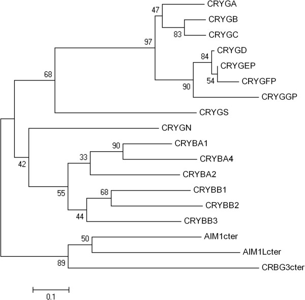
Phylogenetic tree of the humanβγ-crystallin gene superfamily. Sequences were extracted from the UCSC web browser. Translated ORFs were aligned and neighbor-joining trees were constructed as in Figure 2. AIM1, AIML, and CRYBG3 contain internal repeats corresponding to three β-crystallin-like genes in addition to regions not related to crystallin genes. For simplicity, the third, most highly conserved crystallin repeat from each gene was used for this alignment (designated AIM1cter, etc.).

Clustering of crystallin genes. Gaps, indicated by approximate sizes, contain non-crystallin genes. Gene orientations are indicated with arrows (dotted for pseudogenes).
Similar articles
-
sHSP in the eye lens: crystallin mutations, cataract and proteostasis.Int J Biochem Cell Biol. 2012 Oct;44(10):1687-97. doi: 10.1016/j.biocel.2012.02.015. Epub 2012 Mar 2. Int J Biochem Cell Biol. 2012. PMID: 22405853 Review.
-
Differential role of arginine mutations on the structure and functions of α-crystallin.Biochim Biophys Acta. 2016 Jan;1860(1 Pt B):199-210. doi: 10.1016/j.bbagen.2015.06.004. Epub 2015 Jun 14. Biochim Biophys Acta. 2016. PMID: 26080000 Free PMC article. Review.
-
Probing the changes in gene expression due to α-crystallin mutations in mouse models of hereditary human cataract.PLoS One. 2018 Jan 16;13(1):e0190817. doi: 10.1371/journal.pone.0190817. eCollection 2018. PLoS One. 2018. PMID: 29338044 Free PMC article.
-
Effects of alpha-crystallin on lens cell function and cataract pathology.Curr Mol Med. 2009 Sep;9(7):887-92. doi: 10.2174/156652409789105598. Curr Mol Med. 2009. PMID: 19860667 Review.
-
Evolutionary Origins of Pax6 Control of Crystallin Genes.Genome Biol Evol. 2017 Aug 1;9(8):2075-2092. doi: 10.1093/gbe/evx153. Genome Biol Evol. 2017. PMID: 28903537 Free PMC article.
Cited by
-
Evolution of crystallins for a role in the vertebrate eye lens.Protein Sci. 2013 Apr;22(4):367-80. doi: 10.1002/pro.2229. Epub 2013 Feb 26. Protein Sci. 2013. PMID: 23389822 Free PMC article. Review.
-
Divalent Cations and the Divergence of βγ-Crystallin Function.Biochemistry. 2019 Nov 12;58(45):4505-4518. doi: 10.1021/acs.biochem.9b00507. Epub 2019 Nov 1. Biochemistry. 2019. PMID: 31647219 Free PMC article.
-
Whole Exome Sequencing of 20 Spanish Families: Candidate Genes for Non-Syndromic Pediatric Cataracts.Int J Mol Sci. 2023 Jul 13;24(14):11429. doi: 10.3390/ijms241411429. Int J Mol Sci. 2023. PMID: 37511188 Free PMC article.
-
A genomic deletion encompassing CRYBB2-CRYBB2P1 is responsible for autosomal recessive congenital cataracts.Hum Genome Var. 2022 Sep 8;9(1):31. doi: 10.1038/s41439-022-00208-7. Hum Genome Var. 2022. PMID: 36075891 Free PMC article.
-
Enzyme complexity in intermediary metabolism.J Inherit Metab Dis. 2015 Jul;38(4):721-7. doi: 10.1007/s10545-015-9821-0. Epub 2015 Feb 21. J Inherit Metab Dis. 2015. PMID: 25700988 Review.
References
-
- Wistow G, Slingsby C. Structure and evolution of crystallins. The Encyclopedia of the Eye. 2010;2:229–238.
-
- Harding JJ, Crabbe MJC. The lens: development, proteins, metabolism and cataract. Eye. 1984;1B:207–492.
Publication types
MeSH terms
Substances
LinkOut - more resources
Full Text Sources
Research Materials
