

The effects of neck flexion on cerebral potentials evoked by visual, auditory and somatosensory stimuli and focal brain blood flow in related sensory cortices
- PMID: 23199306
- PMCID: PMC3545836
- DOI: 10.1186/1880-6805-31-31
The effects of neck flexion on cerebral potentials evoked by visual, auditory and somatosensory stimuli and focal brain blood flow in related sensory cortices
Abstract
Background: A flexed neck posture leads to non-specific activation of the brain. Sensory evoked cerebral potentials and focal brain blood flow have been used to evaluate the activation of the sensory cortex. We investigated the effects of a flexed neck posture on the cerebral potentials evoked by visual, auditory and somatosensory stimuli and focal brain blood flow in the related sensory cortices.
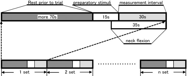
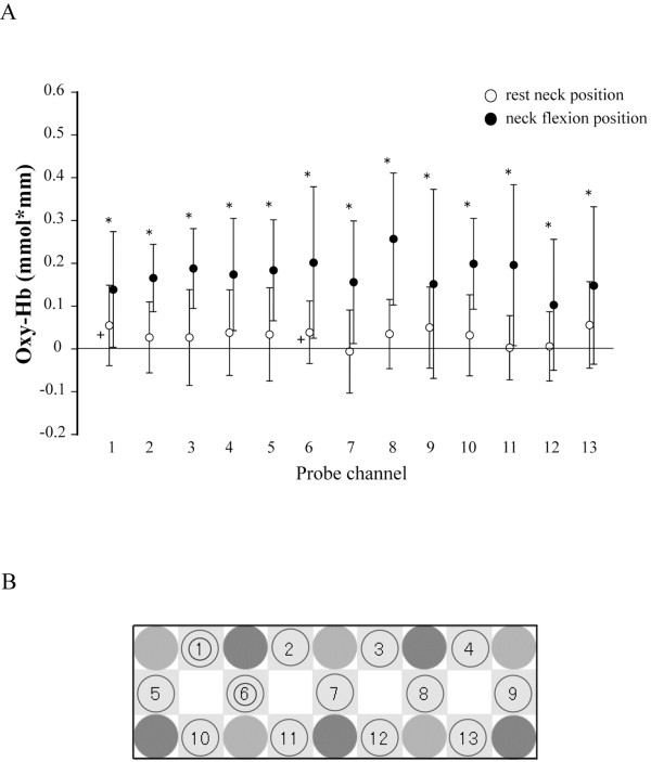
Methods: Twelve healthy young adults received right visual hemi-field, binaural auditory and left median nerve stimuli while sitting with the neck in a resting and flexed (20° flexion) position. Sensory evoked potentials were recorded from the right occipital region, Cz in accordance with the international 10-20 system, and 2 cm posterior from C4, during visual, auditory and somatosensory stimulations. The oxidative-hemoglobin concentration was measured in the respective sensory cortex using near-infrared spectroscopy.
Results: Latencies of the late component of all sensory evoked potentials significantly shortened, and the amplitude of auditory evoked potentials increased when the neck was in a flexed position. Oxidative-hemoglobin concentrations in the left and right visual cortices were higher during visual stimulation in the flexed neck position. The left visual cortex is responsible for receiving the visual information. In addition, oxidative-hemoglobin concentrations in the bilateral auditory cortex during auditory stimulation, and in the right somatosensory cortex during somatosensory stimulation, were higher in the flexed neck position.
Conclusions: Visual, auditory and somatosensory pathways were activated by neck flexion. The sensory cortices were selectively activated, reflecting the modalities in sensory projection to the cerebral cortex and inter-hemispheric connections.
Figures


Similar articles
-
Aging Enhances Neural Activity in Auditory, Visual, and Somatosensory Cortices: The Common Cause Revisited.J Neurosci. 2022 Jan 12;42(2):264-275. doi: 10.1523/JNEUROSCI.0864-21.2021. Epub 2021 Nov 12. J Neurosci. 2022. PMID: 34772740 Free PMC article.
-
Functional impairment due to white matter ischemia after middle cerebral artery occlusion in cats.Stroke. 1990 Jun;21(6):923-8. doi: 10.1161/01.str.21.6.923. Stroke. 1990. PMID: 2349596
-
Developmental and aging changes in somatosensory, auditory and visual evoked potentials.Electroencephalogr Clin Neurophysiol. 1984 Jul;58(1):14-24. doi: 10.1016/0013-4694(84)90196-2. Electroencephalogr Clin Neurophysiol. 1984. PMID: 6203699
-
How is electrical stimulation of the brain experienced, and how can we tell? Selected considerations on sensorimotor function and speech.Cogn Neuropsychol. 2019 May-Jun;36(3-4):103-116. doi: 10.1080/02643294.2019.1609918. Epub 2019 May 10. Cogn Neuropsychol. 2019. PMID: 31076014 Free PMC article. Review.
-
Convergence of Auditory, Visual, and Somatosensory Information in Ventral Prefrontal Cortex.In: Murray MM, Wallace MT, editors. The Neural Bases of Multisensory Processes. Boca Raton (FL): CRC Press/Taylor & Francis; 2012. Chapter 33. In: Murray MM, Wallace MT, editors. The Neural Bases of Multisensory Processes. Boca Raton (FL): CRC Press/Taylor & Francis; 2012. Chapter 33. PMID: 22593866 Free Books & Documents. Review.
Cited by
-
Developmental changes in shortening of pro-saccade reaction time while maintaining neck flexion position.J Physiol Anthropol. 2018 Jan 10;37(1):2. doi: 10.1186/s40101-017-0161-7. J Physiol Anthropol. 2018. PMID: 29321065 Free PMC article.
-
Effect of maintaining neck flexion on anti-saccade reaction time: an investigation using transcranial magnetic stimulation to the frontal oculomotor field.J Physiol Anthropol. 2013 Nov 13;32(1):21. doi: 10.1186/1880-6805-32-21. J Physiol Anthropol. 2013. PMID: 24220550 Free PMC article.
References
MeSH terms
Substances
LinkOut - more resources
Full Text Sources
Medical
Miscellaneous
