

Building a centriole
- PMID: 23199753
- PMCID: PMC3578074
- DOI: 10.1016/j.ceb.2012.10.016
Building a centriole
Abstract
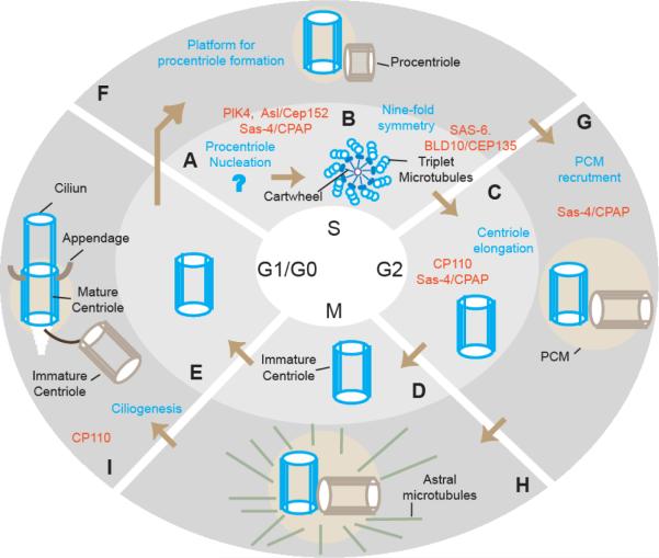
Centrioles are the key foundation of centrosomes and cilia, yet a molecular understanding of how they form has only recently begun to emerge. Building a fully functional centriole that can form a centrosome and cilium requires two cell cycles. Centriole building starts with procentriole nucleation, a process that is coordinated by the conserved proteins Plk4/Zyg-1, and Asterless/Cep152. Subsequently, Sas-6, a conserved procentriole protein, self-assembles to provide nine-fold symmetry to the centriole scaffold. The procentriole then continues to elongate into a centriole, a process controlled by Sas-4/CPAP and CP110. Then, centrioles recruit Sas-4-mediated pre-assembled centrosomal complexes from the cytoplasm to form the pericentriolar material (PCM). Finally, CP110 and its interacting proteins are involved in controlling the timing of centriole templating of the cilium.
Copyright © 2012 Elsevier Ltd. All rights reserved.
Figures
References
-
- Januschke J, Llamazares S, Reina J, Gonzalez C. Drosophila neuroblasts retain the daughter centrosome. Nat Commun. 2011;2(243) - PMC - PubMed
-
* Studying Drosophila neuroblasts using photo converted centrioles and a daughter-centriole-specific markers,the authors show that upon asymmetric mitosis, the old centrosome is inherited by the differentiating daughter cell while the stem cells inherit the new centriole. This demonstrates that old and new centrioles are functionally distinct, but this distinction is used differently from one cell type to the other
-
- Dammermann A, Muller-Reichert T, Pelletier L, Habermann B, Desai A, Oegema K. Centriole assembly requires both centriolar and pericentriolar material proteins. Dev Cell. 2004;7(6):815–829. - PubMed
-
- Pelletier L, O'Toole E, Schwager A, Hyman AA, Muller-Reichert T. Centriole assembly in caenorhabditis elegans. Nature. 2006;444(7119):619–623. - PubMed
-
- Kleylein-Sohn J, Westendorf J, Le Clech M, Habedanck R, Stierhof YD, Nigg EA. Plk4-induced centriole biogenesis in human cells. Dev Cell. 2007;13(2):190–202. - PubMed
Publication types
MeSH terms
Grants and funding
LinkOut - more resources
Full Text Sources
Other Literature Sources
