. 2012 Nov 21;103(10):2071-81.
doi: 10.1016/j.bpj.2012.10.016. Epub 2012 Nov 20.

GABA binding to an insect GABA receptor: a molecular dynamics and mutagenesis study

Affiliations

GABA binding to an insect GABA receptor: a molecular dynamics and mutagenesis study

Jamie A Ashby et al. Biophys J. .

Abstract

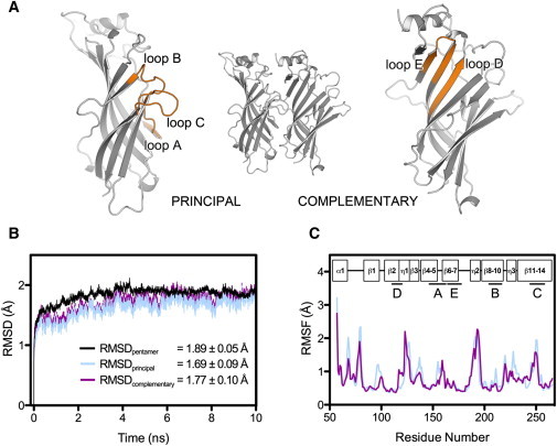
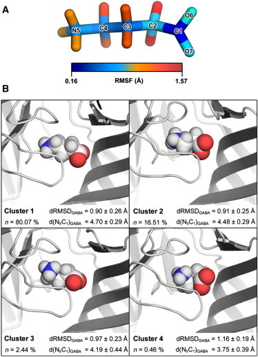
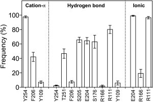
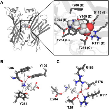
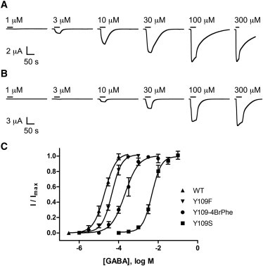
RDL receptors are GABA-activated inhibitory Cys-loop receptors found throughout the insect CNS. They are a key target for insecticides. Here, we characterize the GABA binding site in RDL receptors using computational and electrophysiological techniques. A homology model of the extracellular domain of RDL was generated and GABA docked into the binding site. Molecular dynamics simulations predicted critical GABA binding interactions with aromatic residues F206, Y254, and Y109 and hydrophilic residues E204, S176, R111, R166, S176, and T251. These residues were mutated, expressed in Xenopus oocytes, and their functions assessed using electrophysiology. The data support the binding mechanism provided by the simulations, which predict that GABA forms many interactions with binding site residues, the most significant of which are cation-π interactions with F206 and Y254, H-bonds with E204, S205, R111, S176, T251, and ionic interactions with R111 and E204. These findings clarify the roles of a range of residues in binding GABA in the RDL receptor, and also show that molecular dynamics simulations are a useful tool to identify specific interactions in Cys-loop receptors.


Figures

Figure 1
Figure 1
(A) Cartoon representation of the RDL-ECD dimer containing a single GABA binding site. The secondary structure elements (binding loops A–E) involved in agonist binding in RDL and other Cys-loop receptors are shown in orange. (B) Time evolution of the RMSD of protein backbone atoms for the pentameric RDL-ECD complex and constituent subunits of binding site BS2, during simulation 1. Values given (inset) represent the mean RMSD (± SD) calculated for the RDL-ECD pentamer (black), principal subunit (blue), and complementary subunit (magenta), over the 10 ns production run. (C) RMSF of protein CA atoms for the RDL-ECD principal subunit (blue) and complementary subunit (magenta), of binding-site BS2 following simulation 1. Boxes (inset) represent the secondary structure topology of a single RDL-ECD subunit relative to the residue positions. The locations of the binding regions (AE) involved in GABA interactions are denoted by straight lines and labeled according to panel (A).
Figure 2
Figure 2
(A) Stick representation of GABA with heavy atoms color-coded to depict their RMSF, as calculated from the 10 ns simulation. (B) Cluster analysis of GABA conformations. Individual GABA structures were extracted from each 10 ns simulation trajectory (n = 20) and clustered according to their structural similarity (49). Data from equivalent clusters were pooled and the middle structure of each resultant cluster pool is presented. The degree to which GABA adopts an elongated conformation was determined from the distance between the amino nitrogen atom and carboxylate carbon atom of GABA (d(N5C1)GABA), averaged over equivalent clusters. The relative deviation of cluster members from the starting GABA conformation was determined without prior fitting and the root-square deviation of atom distances (dRMSD) were averaged over equivalent clusters. Statistics refer to the mean ± SD.
Figure 3
Figure 3
Percentage occurrence (± SEM) of interactions between GABA and binding site residues of RDL during the last 8 ns of simulation. Data were averaged over 20 independent binding sites.
Figure 4
Figure 4
(A) Cartoon representation of the RDL-ECD binding site occupied by GABA (left) and the position of GABA interacting amino acid side chains within the binding site (inset). (B) Representative MD snapshot of the interaction between GABA and aromatic residues F206, Y109, and Y254. Gray dashed lines represent cation-π interactions and hydrogen bonds are represented by red dashed lines. Hydrogen bonds were also detected between the carboxylate oxygen atoms of GABA and the OH group of Y254 (not shown). (C) Hydrogen bonds involving nonaromatic amino acids. An additional ionic interaction is formed between the amino moiety of GABA and the carboxylate group of E204.
Figure 5
Figure 5
(A and B) Current traces indicating the response to application of GABA (as denoted by the horizontal black lines) in oocytes expressing WT RDL (A) and Y109F mutant receptors (B). Concentration-response curves from WT RDL and Y109F, Y109S, and Y109-4BrPhe mutants are shown in panel (C). Points represent the mean ± SEM, n = 3–5.

Similar articles

Cited by

References

    1. Thompson A.J., Lester H.A., Lummis S.C.R. The structural basis of function in Cys-loop receptors. Q. Rev. Biophys. 2010;43:449–499. - PubMed
    1. Lummis S.C.R. Locating GABA in GABA receptor binding sites. Biochem. Soc. Trans. 2009;37:1343–1346. - PubMed
    1. Jones M.V., Sahara Y., Westbrook G.L. Defining affinity with the GABAA receptor. J. Neurosci. 1998;18:8590–8604. - PMC - PubMed
    1. McGonigle I., Lummis S.C.R. Molecular characterization of agonists that bind to an insect GABA receptor. Biochemistry. 2010;49:2897–2902. - PMC - PubMed
    1. Woodward R.M., Polenzani L., Miledi R. Characterization of bicuculline/baclofen-insensitive (ρ-like) γ-aminobutyric acid receptors expressed in Xenopus oocytes. II. Pharmacology of γ-aminobutyric acidA and γ-aminobutyric acidB receptor agonists and antagonists. Mol. Pharmacol. 1993;43:609–625. - PubMed

Publication types